1P7D

Crystal structure of the Lambda Integrase (residues 75-356) bound to DNA


Experimental Data Snapshot

  • Method: X-RAY DIFFRACTION
  • Resolution: 2.95 Å
  • R-Value Free: 0.259 
  • R-Value Work: 0.231 

wwPDB Validation   3D Report Full Report


This is version 1.2 of the entry. See complete history


Literature

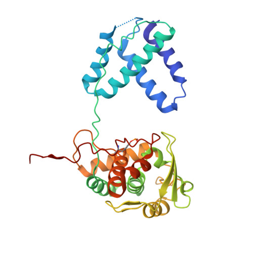
A Conformational Switch Controls the DNA Cleavage Activity of Lambda Integrase

Aihara, H.Kwon, H.J.Nunes-Duby, S.E.Landy, A.Ellenberger, T.

(2003) Mol Cell 12: 187-198

  • DOI: https://doi.org/10.1016/s1097-2765(03)00268-5
  • Primary Citation of Related Structures:  
    1P7D

  • PubMed Abstract: 

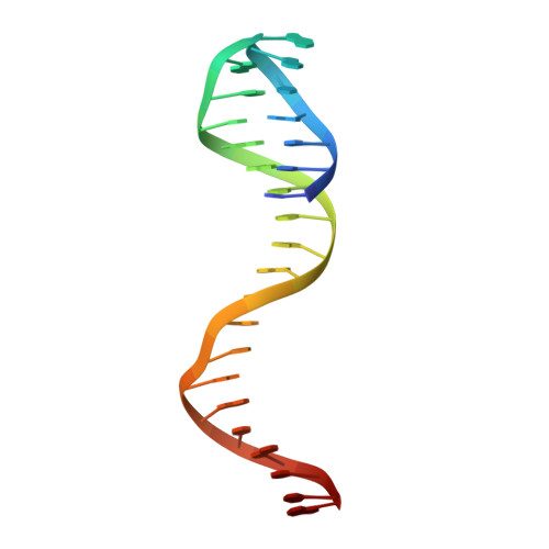
    The bacteriophage lambda integrase protein (lambda Int) belongs to a family of tyrosine recombinases that catalyze DNA rearrangements. We have determined a crystal structure of lambda Int complexed with a cleaved DNA substrate through a covalent phosphotyrosine bond. In comparison to an earlier unliganded structure, we observe a drastic conformational change in DNA-bound lambda Int that brings Tyr342 into the active site for cleavage of the DNA in cis. A flexible linker connects the central and the catalytic domains, allowing the protein to encircle the DNA. Binding specificity is achieved through direct interactions with the DNA and indirect readout of the flexibility of the att site. The conformational switch that activates lambda Int for DNA cleavage exposes the C-terminal 8 residues for interactions with a neighboring Int molecule. The protein interactions mediated by lambda Int's C-terminal tail offer a mechanism for the allosteric control of cleavage activity in higher order lambda Int complexes.


  • Organizational Affiliation

    Department of Biological Chemistry and Molecular Pharmacology, Harvard Medical School, 240 Longwood Avenue, Boston, MA 02115, USA.


Macromolecules

Find similar proteins by:  (by identity cutoff)  |  3D Structure
Entity ID: 3
MoleculeChains Sequence LengthOrganismDetailsImage
IntegraseE [auth A],
F [auth B]
283Lambdavirus lambdaMutation(s): 1 
Gene Names: INT
UniProt
Find proteins for P03700 (Escherichia phage lambda)
Explore P03700 
Go to UniProtKB:  P03700
Entity Groups  
Sequence Clusters30% Identity50% Identity70% Identity90% Identity95% Identity100% Identity
UniProt GroupP03700
Sequence Annotations
Expand
  • Reference Sequence

Find similar nucleic acids by:  Sequence   |   3D Structure  

Entity ID: 1
MoleculeChains LengthOrganismImage
5'-D(*CP*AP*AP*TP*GP*CP*CP*AP*AP*CP*TP*TP*T)-3'A [auth C],
C [auth E]
13N/A
Sequence Annotations
Expand
  • Reference Sequence
Find similar nucleic acids by:  (by identity cutoff)  |  3D Structure
Entity ID: 2
MoleculeChains LengthOrganismImage
26-MERB [auth D],
D [auth F]
26N/A
Sequence Annotations
Expand
  • Reference Sequence
Small Molecules
Modified Residues  1 Unique
IDChains TypeFormula2D DiagramParent
PTR
Query on PTR
E [auth A],
F [auth B]
L-PEPTIDE LINKINGC9 H12 N O6 PTYR
Experimental Data & Validation

Experimental Data

  • Method: X-RAY DIFFRACTION
  • Resolution: 2.95 Å
  • R-Value Free: 0.259 
  • R-Value Work: 0.231 
  • Space Group: P 1 21 1
Unit Cell:
Length ( Å )Angle ( ˚ )
a = 59.021α = 90
b = 106.161β = 92.1
c = 73.165γ = 90
Software Package:
Software NamePurpose
HKL-2000data collection
TRUNCATEdata reduction
MLPHAREphasing
CNSrefinement
HKL-2000data reduction
CCP4data scaling

Structure Validation

View Full Validation Report


Entry History 

Deposition Data

Revision History  (Full details and data files)

  • Version 1.0: 2003-08-12
    Type: Initial release
  • Version 1.1: 2008-04-29
    Changes: Version format compliance
  • Version 1.2: 2011-07-13
    Changes: Version format compliance