1P3H

Crystal Structure of the Mycobacterium tuberculosis chaperonin 10 tetradecamer


Experimental Data Snapshot

  • Method: X-RAY DIFFRACTION
  • Resolution: 2.80 Å
  • R-Value Free: 0.280 
  • R-Value Work: 0.259 
  • R-Value Observed: 0.260 

wwPDB Validation   3D Report Full Report


This is version 1.5 of the entry. See complete history


Literature

Mycobacterium tuberculosis chaperonin 10 heptamers self-associate through their biologically active loops

Roberts, M.M.Coker, A.R.Fossati, G.Mascagni, P.Coates, A.R.M.Wood, S.P.

(2003) J Bacteriol 185: 4172-4185

  • DOI: https://doi.org/10.1128/JB.185.14.4172-4185.2003
  • Primary Citation of Related Structures:  
    1P3H

  • PubMed Abstract: 

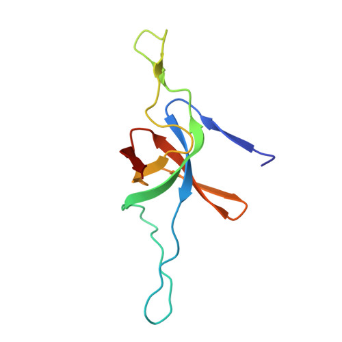
    The crystal structure of Mycobacterium tuberculosis chaperonin 10 (cpn10(Mt)) has been determined to a resolution of 2.8 A. Two dome-shaped cpn10(Mt) heptamers complex through loops at their bases to form a tetradecamer with 72 symmetry and a spherical cage-like structure. The hollow interior enclosed by the tetradecamer is lined with hydrophilic residues and has dimensions of 30 A perpendicular to and 60 A along the sevenfold axis. Tetradecameric cpn10(Mt) has also been observed in solution by dynamic light scattering. Through its base loop sequence cpn10(Mt) is known to be the agent in the bacterium responsible for bone resorption and for the contribution towards its strong T-cell immunogenicity. Superimposition of the cpn10(Mt) sequences 26 to 32 and 66 to 72 and E. coli GroES 25 to 31 associated with bone resorption activity shows them to have similar conformations and structural features, suggesting that there may be a common receptor for the bone resorption sequences. The base loops of cpn10s in general also attach to the corresponding chaperonin 60 (cpn60) to enclose unfolded protein and to facilitate its correct folding in vivo. Electron density corresponding to a partially disordered protein subunit appears encapsulated within the interior dome cavity of each heptamer. This suggests that the binding of substrates to cpn10 is possible in the absence of cpn60.


  • Organizational Affiliation

    Medical Microbiology, Department of Cellular and Molecular Medicine, St. George's Hospital Medical School, London SW17 0RE, England. m.roberts@sghms.ac.uk


Macromolecules
Find similar proteins by:  (by identity cutoff)  |  3D Structure
Entity ID: 1
MoleculeChains Sequence LengthOrganismDetailsImage
10 kDa chaperonin
A, B, C, D, E
A, B, C, D, E, F, G, H, I, J, K, L, M, N
99Mycobacterium tuberculosisMutation(s): 0 
Gene Names: GROS OR GROES OR MOPB OR CPN10 OR RV3418C OR MT3527 OR MTCY78.11
UniProt
Find proteins for P9WPE5 (Mycobacterium tuberculosis (strain ATCC 25618 / H37Rv))
Explore P9WPE5 
Go to UniProtKB:  P9WPE5
Entity Groups  
Sequence Clusters30% Identity50% Identity70% Identity90% Identity95% Identity100% Identity
UniProt GroupP9WPE5
Sequence Annotations
Expand
  • Reference Sequence
Small Molecules
Ligands 2 Unique
IDChains Name / Formula / InChI Key2D Diagram3D Interactions
MPD
Query on MPD

Download Ideal Coordinates CCD File 
AA [auth H]
BA [auth I]
CA [auth J]
DA [auth K]
EA [auth L]
AA [auth H],
BA [auth I],
CA [auth J],
DA [auth K],
EA [auth L],
FA [auth M],
GA [auth N],
P [auth A],
Q [auth A],
R [auth B],
S [auth C],
T [auth D],
U [auth E],
V [auth F],
W [auth G],
X [auth H],
Y [auth H],
Z [auth H]
(4S)-2-METHYL-2,4-PENTANEDIOL
C6 H14 O2
SVTBMSDMJJWYQN-YFKPBYRVSA-N
CA
Query on CA

Download Ideal Coordinates CCD File 
O [auth A]CALCIUM ION
Ca
BHPQYMZQTOCNFJ-UHFFFAOYSA-N
Experimental Data & Validation

Experimental Data

  • Method: X-RAY DIFFRACTION
  • Resolution: 2.80 Å
  • R-Value Free: 0.280 
  • R-Value Work: 0.259 
  • R-Value Observed: 0.260 
  • Space Group: P 1 21 1
Unit Cell:
Length ( Å )Angle ( ˚ )
a = 76.46α = 90
b = 87.93β = 106.78
c = 124.39γ = 90
Software Package:
Software NamePurpose
XDSdata scaling
TRUNCATEdata reduction
DMmodel building
CNSrefinement
XDSdata reduction
CCP4data scaling
DMphasing

Structure Validation

View Full Validation Report


Entry History 

Deposition Data

Revision History  (Full details and data files)

  • Version 1.0: 2003-07-15
    Type: Initial release
  • Version 1.1: 2008-04-29
    Changes: Version format compliance
  • Version 1.2: 2011-07-13
    Changes: Version format compliance
  • Version 1.3: 2017-10-11
    Changes: Advisory, Refinement description
  • Version 1.4: 2024-02-14
    Changes: Advisory, Data collection, Database references, Derived calculations
  • Version 1.5: 2024-04-03
    Changes: Refinement description