1OUB

CONTRIBUTION OF HYDROPHOBIC RESIDUES TO THE STABILITY OF HUMAN LYSOZYME: X-RAY STRUCTURE OF THE V100A MUTANT


Experimental Data Snapshot

  • Method: X-RAY DIFFRACTION
  • Resolution: 1.80 Å
  • R-Value Work: 0.160 
  • R-Value Observed: 0.160 

wwPDB Validation   3D Report Full Report


This is version 1.4 of the entry. See complete history


Literature

Contribution of the hydrophobic effect to the stability of human lysozyme: calorimetric studies and X-ray structural analyses of the nine valine to alanine mutants.

Takano, K.Yamagata, Y.Fujii, S.Yutani, K.

(1997) Biochemistry 36: 688-698

  • DOI: https://doi.org/10.1021/bi9621829
  • Primary Citation of Related Structures:  
    1OUB, 1OUC, 1OUD, 1OUE, 1OUF, 1OUG, 1OUH, 1OUI, 1OUJ

  • PubMed Abstract: 

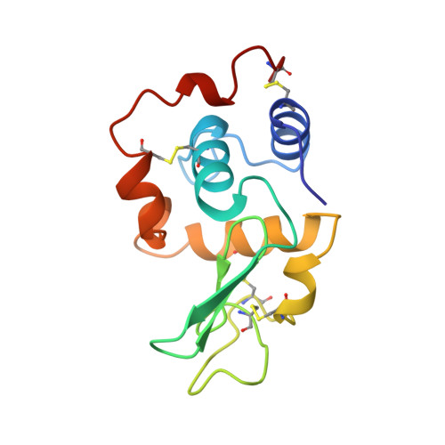
    To clarify the contribution of the hydrophobic effect to the conformational stability of human lysozyme, a series of Val to Ala mutants were constructed. The thermodynamic parameters for the denaturation of these nine mutant proteins were determined using differential scanning calorimetry (DSC), and the crystal structures were solved at high resolution. The denaturation Gibbs energy (delta delta G) and enthalpy (delta delta H) values of the mutant proteins ranged from +2.2 to- 6.3 kJ/mol and from +7 to -17 kJ/mol, respectively. The structural analyses showed that the mutation site and/or the residues around it in some proteins shifted toward the created cavity, and the substitutions affected not only the mutations site but also other parts far from the site, although the structural changes were not as great. Correlation between the changes in the thermodynamic parameters and the structural features of mutant proteins was examined, including the five Ile to Val mutant human lysozymes [Takano et al. (1995) J. Mol. Biol. 254, 62-76]. There was no simple general correlation between delta delta G and the changes in hydrophobic surface area exposed upon denaturation (delta delta ASAHP). We found only a new correlation between the delta delta G and delta delta ASAHP of all of the hydrophobic residues if the effect of the secondary structure propensity was taken into account.


  • Organizational Affiliation

    Institute for Protein Research, Osaka University, Japan.


Macromolecules
Find similar proteins by:  (by identity cutoff)  |  3D Structure
Entity ID: 1
MoleculeChains Sequence LengthOrganismDetailsImage
LYSOZYME130Homo sapiensMutation(s): 1 
Gene Names: HUMAN LYSOZYME WITH VAL 100
EC: 3.2.1.17
UniProt & NIH Common Fund Data Resources
Find proteins for P61626 (Homo sapiens)
Explore P61626 
Go to UniProtKB:  P61626
PHAROS:  P61626
GTEx:  ENSG00000090382 
Entity Groups  
Sequence Clusters30% Identity50% Identity70% Identity90% Identity95% Identity100% Identity
UniProt GroupP61626
Sequence Annotations
Expand
  • Reference Sequence
Small Molecules
Ligands 1 Unique
IDChains Name / Formula / InChI Key2D Diagram3D Interactions
NA
Query on NA

Download Ideal Coordinates CCD File 
B [auth A]SODIUM ION
Na
FKNQFGJONOIPTF-UHFFFAOYSA-N
Experimental Data & Validation

Experimental Data

  • Method: X-RAY DIFFRACTION
  • Resolution: 1.80 Å
  • R-Value Work: 0.160 
  • R-Value Observed: 0.160 
  • Space Group: P 21 21 21
Unit Cell:
Length ( Å )Angle ( ˚ )
a = 56.56α = 90
b = 61β = 90
c = 33.7γ = 90
Software Package:
Software NamePurpose
RIGAKUdata collection
RIGAKUdata reduction
X-PLORmodel building
X-PLORrefinement
RIGAKUdata scaling
X-PLORphasing

Structure Validation

View Full Validation Report


Entry History 

Deposition Data

Revision History  (Full details and data files)

  • Version 1.0: 1997-02-12
    Type: Initial release
  • Version 1.1: 2008-03-24
    Changes: Version format compliance
  • Version 1.2: 2011-07-13
    Changes: Version format compliance
  • Version 1.3: 2017-11-29
    Changes: Derived calculations, Other
  • Version 1.4: 2021-11-03
    Changes: Database references, Derived calculations