N2-Substituted O6-Cyclohexylmethylguanine Derivatives: Potent Inhibitors of Cyclin-Dependent Kinases 1 and 2
Hardcastle, I.R., Arris, C.E., Bentley, J., Boyle, F.T., Chen, Y., Curtin, N.J., Endicott, J.A., Gibson, A.E., Golding, B.T., Griffin, R.J., Jewsbury, P., Menyerol, J., Mesguiche, V., Newell, D.R., Noble, M.E.M., Pratt, D.J., Wang, L.-Z., Whitfield, H.J.(2004) J Med Chem 47: 3710
- PubMed: 15239650 Search on PubMed
- DOI: https://doi.org/10.1021/jm0311442
- Primary Citation Related Structures:
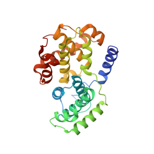
1OI9, 1OIU, 1OIY - PubMed Abstract:
The adenosine 5'-triphosphate (ATP) competitive cyclin-dependent kinase inhibitor O(6)-cyclohexylmethylguanine (NU2058, 1) has been employed as the lead in a structure-based drug discovery program resulting in the discovery of the potent CDK1 and -2 inhibitor NU6102 (3, IC(50) = 9.5 nM and 5.4 nM vs CDK1/cyclinB and CDK2/cyclinA3, respectively). The SAR for this series have been explored further by the synthesis and evaluation of 45 N(2)-substituted analogues of NU2058. These studies have confirmed the requirement for the hydrogen bonding N(2)-NH group and the requirement for an aromatic N(2)-substituent to confer potency in the series. Additional potency is conferred by the presence of a group capable of donating a hydrogen bond at the 4'-position, for example, the 4'-hydroxy derivative (25, IC(50) = 94 nM and 69 nM vs CDK1/cyclinB and CDK2/cyclinA3, respectively), 4'-monomethylsulfonamide derivative (28, IC(50) = 9 nM and 7.0 nM vs CDK1/cyclinB and CDK2/cyclinA3, respectively), and 4'-carboxamide derivative (34, IC(50) = 67 nM and 64 nM vs CDK1/cyclinB and CDK2/cyclinA3, respectively). X-ray crystal structures have been obtained for key compounds and have been used to explain the observed trends in activity.
- Northern Institute for Cancer Research, Bedson Building, School of Natural Sciences, University of Newcastle, Newcastle upon Tyne NE1 7RU, UK. I.R.Hardcastle@ncl.ac.uk
Organizational Affiliation: