1OEM | pdb_00001oem

PTP1B with the catalytic cysteine oxidized to a sulfenyl-amide bond


Experimental Data Snapshot

  • Method: X-RAY DIFFRACTION
  • Resolution: 1.80 Å
  • R-Value Free: 
    0.227 (Depositor), 0.215 (DCC) 
  • R-Value Work: 
    0.203 (Depositor), 0.203 (DCC) 
  • R-Value Observed: 
    0.204 (Depositor) 

Starting Model: experimental
View more details

wwPDB Validation 3D Report Full Report

Validation slider image for 1OEM

This is version 1.3 of the entry. See complete history

Literature

Redox Regulation of Protein Tyrosine Phosphatase Involves a Sulfenyl-Amide Intermediate

Salmeen, A.Andersen, J.N.Myers, M.P.Meng, T.C.Hinks, J.A.Tonks, N.K.Barford, D.

(2003) Nature 423: 769

  • DOI: https://doi.org/10.1038/nature01680
  • Primary Citation Related Structures: 
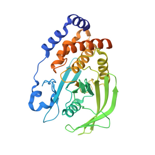
    1OEM, 1OEO

  • PubMed Abstract: 

    The second messenger hydrogen peroxide is required for optimal activation of numerous signal transduction pathways, particularly those mediated by protein tyrosine kinases. One mechanism by which hydrogen peroxide regulates cellular processes is the transient inhibition of protein tyrosine phosphatases through the reversible oxidization of their catalytic cysteine, which suppresses protein dephosphorylation. Here we describe a structural analysis of the redox-dependent regulation of protein tyrosine phosphatase 1B (PTP1B), which is reversibly inhibited by oxidation after cells are stimulated with insulin and epidermal growth factor. The sulphenic acid intermediate produced in response to PTP1B oxidation is rapidly converted into a previously unknown sulphenyl-amide species, in which the sulphur atom of the catalytic cysteine is covalently linked to the main chain nitrogen of an adjacent residue. Oxidation of PTP1B to the sulphenyl-amide form is accompanied by large conformational changes in the catalytic site that inhibit substrate binding. We propose that this unusual protein modification both protects the active-site cysteine residue of PTP1B from irreversible oxidation to sulphonic acid and permits redox regulation of the enzyme by promoting its reversible reduction by thiols.


  • Organizational Affiliation
    • Section of Structural Biology, Institute of Cancer Research, Chester Beatty Laboratories, 237 Fulham Road, London SW3 6JB, UK.

Macromolecule Content 

  • Total Structure Weight: 37.37 kDa 
  • Atom Count: 2,470 
  • Modeled Residue Count: 281 
  • Deposited Residue Count: 321 
  • Unique protein chains: 1

Macromolecules

Find similar proteins by:|  3D Structure
Entity ID: 1
MoleculeChains  Sequence LengthOrganismDetailsImage
PROTEIN-TYROSINE PHOSPHATASE, NON-RECEPTOR TYPE 1A [auth X]321Homo sapiensMutation(s): 0 
EC: 3.1.3.48
UniProt & NIH Common Fund Data Resources
Find proteins for P18031 (Homo sapiens)
Explore P18031 
Go to UniProtKB:  P18031
PHAROS:  P18031
GTEx:  ENSG00000196396 
Entity Groups
Sequence Clusters30% Identity50% Identity70% Identity90% Identity95% Identity100% Identity
UniProt GroupP18031
Sequence Annotations
Expand
Reference Sequence

Experimental Data & Validation

Experimental Data

  • Method: X-RAY DIFFRACTION
  • Resolution: 1.80 Å
  • R-Value Free:  0.227 (Depositor), 0.215 (DCC) 
  • R-Value Work:  0.203 (Depositor), 0.203 (DCC) 
  • R-Value Observed: 0.204 (Depositor) 
Space Group: P 31 2 1
Unit Cell:
Length ( Å )Angle ( ˚ )
a = 88.218α = 90
b = 88.218β = 90
c = 103.787γ = 120
Software Package:
Software NamePurpose
REFMACrefinement
DENZOdata reduction
SCALEPACKdata scaling
AMoREphasing

Structure Validation

View Full Validation Report


Entry History 

Deposition Data

Revision History  (Full details and data files)

  • Version 1.0: 2003-06-12
    Type: Initial release
  • Version 1.1: 2011-05-08
    Changes: Version format compliance
  • Version 1.2: 2011-07-13
    Changes: Version format compliance
  • Version 1.3: 2023-12-13
    Changes: Data collection, Database references, Refinement description