1OCY

Structure of the receptor-binding domain of the bacteriophage T4 short tail fibre


Experimental Data Snapshot

  • Method: X-RAY DIFFRACTION
  • Resolution: 1.50 Å
  • R-Value Free: 0.152 
  • R-Value Work: 0.143 

wwPDB Validation   3D Report Full Report


This is version 1.3 of the entry. See complete history


Literature

The Structure of the Receptor-Binding Domain of the Bacteriophage T4 Short Tail Fibre Reveals a Knitted Trimeric Metal-Binding Fold

Thomassen, E.Gielen, G.Schuetz, M.Schoehn, G.Abrahams, J.P.Miller, S.van Raaij, M.J.

(2003) J Mol Biol 331: 361-373

  • DOI: https://doi.org/10.1016/s0022-2836(03)00755-1
  • Primary Citation of Related Structures:  
    1OCY

  • PubMed Abstract: 

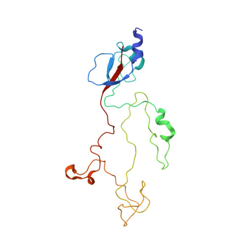
    Adsorption of T4 bacteriophage to the Escherichia coli host cell is mediated by six long and six short tail fibres. After at least three long tail fibres have bound, short tail fibres extend and bind irreversibly to the core region of the host cell lipo-polysaccharide (LPS), serving as inextensible stays during penetration of the cell envelope by the tail tube. The short tail fibres consist of a parallel, in-register, trimer of gene product 12 (gp12).X-ray crystallography at 1.5A resolution of a protease-stable fragment of gp12 generated in the presence of zinc chloride reveals the structure of the C-terminal receptor-binding domain. It has a novel "knitted" fold, consisting of three extensively intertwined monomers. It reveals a metal-binding site, containing a zinc ion coordinated by six histidine residues in an octahedral conformation. We also suggest an LPS-binding region.


  • Organizational Affiliation

    Biophysical Structural Chemistry, Leiden Institute of Chemistry, Leiden University, Einsteinweg 55, NL-2333 CC, Leiden, The Netherlands.


Macromolecules
Find similar proteins by:  (by identity cutoff)  |  3D Structure
Entity ID: 1
MoleculeChains Sequence LengthOrganismDetailsImage
BACTERIOPHAGE T4 SHORT TAIL FIBRE198Tequatrovirus T4Mutation(s): 0 
UniProt
Find proteins for P10930 (Enterobacteria phage T4)
Explore P10930 
Go to UniProtKB:  P10930
Entity Groups  
Sequence Clusters30% Identity50% Identity70% Identity90% Identity95% Identity100% Identity
UniProt GroupP10930
Sequence Annotations
Expand
  • Reference Sequence
Experimental Data & Validation

Experimental Data

  • Method: X-RAY DIFFRACTION
  • Resolution: 1.50 Å
  • R-Value Free: 0.152 
  • R-Value Work: 0.143 
  • Space Group: P 3 2 1
Unit Cell:
Length ( Å )Angle ( ˚ )
a = 50.782α = 90
b = 50.782β = 90
c = 435.647γ = 120
Software Package:
Software NamePurpose
REFMACrefinement
MOSFLMdata reduction
SCALAdata scaling
SOLVE/RESOLVEphasing

Structure Validation

View Full Validation Report


Entry History 

Deposition Data

Revision History  (Full details and data files)

  • Version 1.0: 2003-07-24
    Type: Initial release
  • Version 1.1: 2011-05-08
    Changes: Version format compliance
  • Version 1.2: 2011-07-13
    Changes: Version format compliance
  • Version 1.3: 2018-02-07
    Changes: Database references, Structure summary