The closed state of a H+ channel helical bundle combining precise orientational and distance restraints from solid state NMR
Nishimura, K., Kim, S., Zhang, L., Cross, T.A.(2002) Biochemistry 41: 13170-13177
- PubMed: 12403618 Search on PubMed
- DOI: https://doi.org/10.1021/bi0262799
- Primary Citation Related Structures:
1NYJ - PubMed Abstract:
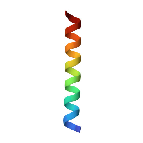
An interhelical distance has been precisely measured by REDOR solid-state NMR spectroscopy in the transmembrane tetrameric bundle of M2-TMP, from the M2 proton channel of the influenza A viral coat. The high-resolution structure of the helical backbone has been determined using orientational restraints from uniformly aligned peptide preparations in hydrated dimyristoylphosphatidylcholine bilayers. Here, the distance between (15)N(pi) labeled His37 and (13)C(gamma) labeled Trp41 is determined to be less than 3.9 A. Such a short distance, in combination with the known tilt and rotational orientation of the individual helices, permits not only a determination of which specific side chain pairings give rise to the interaction, but also the side chain torsion angles and restraints for the tetrameric bundle can also be characterized. The resulting proton channel structure is validated in a variety of ways. Both histidine and tryptophan side chains are oriented in toward the pore where they can play a significant functional role. The channel appears to be closed by the proximity of the four indoles consistent with electrophysiology and mutagenesis studies of the intact protein at pH 7.0 and above. The pore maintains its integrity to the N terminal side of the membrane, and at the same time, a cavity is generated that appears adequate for binding amantadine. Finally, the observation of a 2 kHz coupling in the PISEMA spectrum of (15)N(pi)His37 validates the orientation of the His37 side chain based on the observed REDOR distance.
- National High Magnetic Field Laboratory, Florida State University, Tallahassee, Florida 32310, USA.
Organizational Affiliation: