1NUB | pdb_00001nub

HELIX C DELETION MUTANT OF BM-40 FS-EC DOMAIN PAIR


Experimental Data Snapshot

  • Method: X-RAY DIFFRACTION
  • Resolution: 2.80 Å
  • R-Value Free: 
    0.306 (Depositor), 0.292 (DCC) 
  • R-Value Work: 
    0.255 (Depositor), 0.245 (DCC) 
  • R-Value Observed: 
    0.255 (Depositor) 

Starting Model: experimental
View more details

wwPDB Validation 3D Report Full Report

Validation slider image for 1NUB

This is version 2.2 of the entry. See complete history

Literature

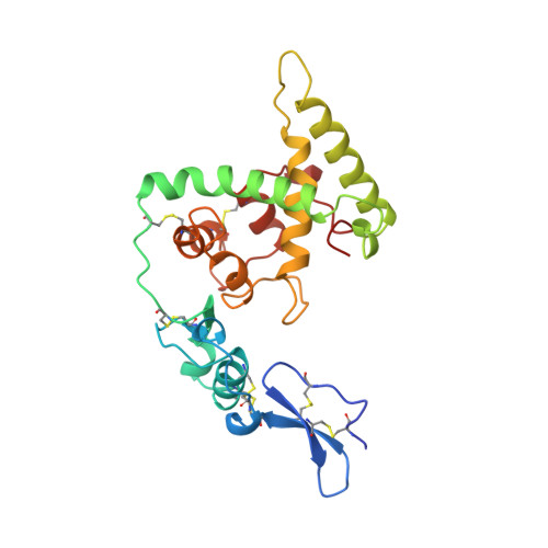
Crystal structure and mapping by site-directed mutagenesis of the collagen-binding epitope of an activated form of BM-40/SPARC/osteonectin.

Sasaki, T.Hohenester, E.Gohring, W.Timpl, R.

(1998) EMBO J 17: 1625-1634

  • DOI: https://doi.org/10.1093/emboj/17.6.1625
  • Primary Citation Related Structures: 
    1NUB

  • PubMed Abstract: 

    The extracellular calcium-binding domain (positions 138-286) of the matrix protein BM-40 possesses a binding epitope of moderate affinity for several collagen types. This epitope was predicted to reside in helix alphaA and to be partially masked by helix alphaC. Here we show that deletion of helix alphaC produces a 10-fold increase in collagen affinity similar to that seen after proteolytic cleavage of this helix. The predicted removal of the steric constraint was clearly demonstrated by the crystal structure of the mutant at 2.8 A resolution. This constitutively activated mutant was used to map the collagen-binding site following alanine mutagenesis at 13 positions. Five residues were crucial for binding, R149 and N156 in helix alphaA, and L242, M245 and E246 in a loop region connecting the two EF hands of BM-40. These residues are spatially close and form a flat ring of 15 A diameter which matches the diameter of a triple-helical collagen domain. The mutations showed similar effects on binding to collagens I and IV, indicating nearly identical binding sites on both collagens. Selected mutations in the non-activated mutant DeltaI also reduced collagen binding, consistent with the same location of the epitope but in a more cryptic form in intact BM-40.


  • Organizational Affiliation
    • Max-Planck-Institut für Biochemie, D-82152, Martinsried, Germany.

Macromolecule Content 

  • Total Structure Weight: 54.13 kDa 
  • Atom Count: 3,732 
  • Modeled Residue Count: 452 
  • Deposited Residue Count: 458 
  • Unique protein chains: 1

Macromolecules

Find similar proteins by:|  3D Structure
Entity ID: 1
MoleculeChains  Sequence LengthOrganismDetailsImage
BASEMENT MEMBRANE PROTEIN BM-40
A, B
229Homo sapiensMutation(s): 0 
UniProt & NIH Common Fund Data Resources
Find proteins for P09486 (Homo sapiens)
Explore P09486 
Go to UniProtKB:  P09486
PHAROS:  P09486
GTEx:  ENSG00000113140 
Entity Groups
Sequence Clusters30% Identity50% Identity70% Identity90% Identity95% Identity100% Identity
UniProt GroupP09486
Glycosylation
Glycosylation Sites: 1Go to GlyGen: P09486-1
Sequence Annotations
Expand
Reference Sequence

Oligosaccharides

Help  
Entity ID: 2
MoleculeChains Length2D Diagram GlycosylationD Interactions
2-acetamido-2-deoxy-beta-D-glucopyranose-(1-4)-2-acetamido-2-deoxy-beta-D-glucopyranose
C, D
2N-Glycosylation
Glycosylation Resources
GlyTouCan: G42666HT
GlyCosmos: G42666HT
GlyGen: G42666HT

Experimental Data & Validation

Experimental Data

  • Method: X-RAY DIFFRACTION
  • Resolution: 2.80 Å
  • R-Value Free:  0.306 (Depositor), 0.292 (DCC) 
  • R-Value Work:  0.255 (Depositor), 0.245 (DCC) 
  • R-Value Observed: 0.255 (Depositor) 
Space Group: P 21 21 21
Unit Cell:
Length ( Å )Angle ( ˚ )
a = 51.86α = 90
b = 88.14β = 90
c = 153.73γ = 90
Software Package:
Software NamePurpose
MOSFLMdata reduction
SCALAdata scaling
X-PLORmodel building
X-PLORrefinement
CCP4data scaling
X-PLORphasing

Structure Validation

View Full Validation Report


Entry History 

Deposition Data

Revision History  (Full details and data files)

  • Version 1.0: 1998-12-30
    Type: Initial release
  • Version 1.1: 2008-03-24
    Changes: Version format compliance
  • Version 1.2: 2011-07-13
    Changes: Non-polymer description, Version format compliance
  • Version 2.0: 2020-07-29
    Type: Remediation
    Reason: Carbohydrate remediation
    Changes: Advisory, Atomic model, Data collection, Database references, Derived calculations, Structure summary
  • Version 2.1: 2024-04-03
    Changes: Advisory, Data collection, Database references, Refinement description, Structure summary
  • Version 2.2: 2024-11-13
    Changes: Structure summary