1NFO | pdb_00001nfo

APOLIPOPROTEIN E2 (APOE2, D154A MUTATION)


Experimental Data Snapshot

  • Method: X-RAY DIFFRACTION
  • Resolution: 2.00 Å
  • R-Value Free: 
    0.270 (Depositor) 
  • R-Value Work: 
    0.232 (DCC) 
  • R-Value Observed: 
    0.206 (Depositor) 

Starting Model: experimental
View more details

wwPDB Validation 3D Report Full Report

Validation slider image for 1NFO

This is version 1.5 of the entry. See complete history

Literature

Novel mechanism for defective receptor binding of apolipoprotein E2 in type III hyperlipoproteinemia.

Dong, L.M.Parkin, S.Trakhanov, S.D.Rupp, B.Simmons, T.Arnold, K.S.Newhouse, Y.M.Innerarity, T.L.Weisgraber, K.H.

(1996) Nat Struct Biol 3: 718-722

  • DOI: https://doi.org/10.1038/nsb0896-718
  • Primary Citation Related Structures: 
    1NFN, 1NFO

  • PubMed Abstract: 

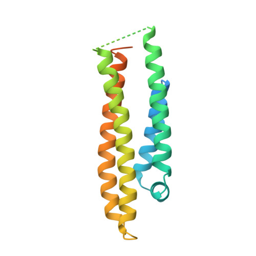
    The defective binding of apolipoprotein (apo) E2 to lipoprotein receptors, an underlying cause of type III hyperlipoproteinemia, results from replacement of Arg 158 with Cys, disrupting the naturally occurring salt bridge between Asp 154 and Arg 158. A new bond between Asp 154 and Arg 150 is formed, shifting Arg 150 out of the receptor binding region. Elimination of the 154-150 salt bridge by site-directed mutagenesis of Asp 154 to Ala restored the receptor binding activity to near normal levels. The X-ray crystal structure of apoE2 Ala 154 demonstrated that Arg 150 was relocated within the receptor binding region. Our results demonstrate that defective binding of apoE2 occurs by a novel mechanism of the replacement of one salt bridge with another.


  • Organizational Affiliation
    • Gladstone Institute of Cardiovascular Disease, University of California, San Francisco 94141-9100, USA.

Macromolecule Content 

  • Total Structure Weight: 22.06 kDa 
  • Atom Count: 1,216 
  • Modeled Residue Count: 131 
  • Deposited Residue Count: 191 
  • Unique protein chains: 1

Macromolecules

Find similar proteins by:|  3D Structure
Entity ID: 1
MoleculeChains  Sequence LengthOrganismDetailsImage
APOLIPOPROTEIN E2191Homo sapiensMutation(s): 1 
Gene Names: E2
UniProt & NIH Common Fund Data Resources
Find proteins for P02649 (Homo sapiens)
Explore P02649 
Go to UniProtKB:  P02649
PHAROS:  P02649
GTEx:  ENSG00000130203 
Entity Groups
Sequence Clusters30% Identity50% Identity70% Identity90% Identity95% Identity100% Identity
UniProt GroupP02649
Sequence Annotations
Expand
Reference Sequence

Experimental Data & Validation

Experimental Data

  • Method: X-RAY DIFFRACTION
  • Resolution: 2.00 Å
  • R-Value Free:  0.270 (Depositor) 
  • R-Value Work:  0.232 (DCC) 
  • R-Value Observed: 0.206 (Depositor) 
Space Group: P 21 21 21
Unit Cell:
Length ( Å )Angle ( ˚ )
a = 40.613α = 90
b = 53.671β = 90
c = 84.795γ = 90
Software Package:
Software NamePurpose
SHELXL-93model building
X-PLORmodel building
SHELXL-93refinement
X-PLORrefinement
ADSCdata collection
SHELXL-93phasing
X-PLORphasing

Structure Validation

View Full Validation Report


Entry History 

Deposition Data

Revision History  (Full details and data files)

  • Version 1.0: 1997-01-27
    Type: Initial release
  • Version 1.1: 2008-03-24
    Changes: Version format compliance
  • Version 1.2: 2011-07-13
    Changes: Version format compliance
  • Version 1.3: 2021-11-03
    Changes: Database references
  • Version 1.4: 2023-08-09
    Changes: Refinement description
  • Version 1.5: 2024-05-22
    Changes: Data collection