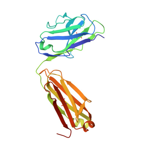
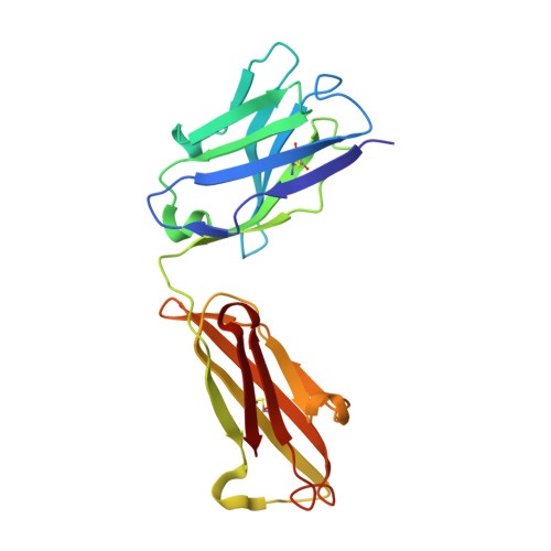
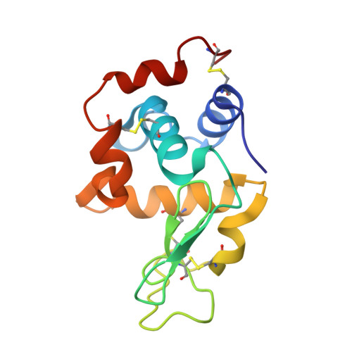
Dissection of binding interactions in the complex between the anti-lysozyme antibody HyHEL-63 and its antigen
Li, Y., Urrutia, M., Smith-Gill, S.J., Mariuzza, R.A.(2003) Biochemistry 42: 11-22
- PubMed: 12515535 Search on PubMed
- DOI: https://doi.org/10.1021/bi020589+
- Primary Citation Related Structures:
1NBY, 1NBZ - PubMed Abstract:
Alanine-scanning mutagenesis, X-ray crystallography, and double mutant cycles were used to characterize the interface between the anti-hen egg white lysozyme (HEL) antibody HyHEL-63 and HEL. Eleven HEL residues in contact with HyHEL-63 in the crystal structure of the antigen-antibody complex, and 10 HyHEL-63 residues in contact with HEL, were individually truncated to alanine in order to determine their relative contributions to complex stabilization. The residues of HEL (Tyr20, Lys96, and Lys97) most important for binding HyHEL-63 (Delta G(mutant) - Delta G(wild type) > 3.0 kcal/mol) form a contiguous patch at the center of the surface contacted by the antibody. Hot spot residues of the antibody (Delta Delta G > 2.0 kcal/mol) are organized in two clusters that juxtapose hot spot residues of HEL, resulting in energetic complementarity across the interface. All energetically critical residues are centrally located, shielded from solvent by peripheral residues that contribute significantly less to the binding free energy. Although HEL hot spot residues Lys96 and Lys97 make similar interactions with antibody in the HyHEL-63/HEL complex, alanine substitution of Lys96 results in a nearly 100-fold greater reduction in affinity than the corresponding mutation in Lys97. To understand the basis for this marked difference, we determined the crystal structures of the HyHEL-63/HEL Lys96Ala and HyHEL-63/HEL Lys97Ala complexes to 1.80 and 1.85 A resolution, respectively. Whereas conformational changes in the proteins and differences in the solvent networks at the mutation sites appear too small to explain the observed affinity difference, superposition of free HEL in different crystal forms onto bound HEL in the wild type and mutant HyHEL-63/HEL complexes reveals that the side-chain conformation of Lys96 is very similar in the various structures, but that the Lys97 side chain displays considerable flexibility. Accordingly, a greater entropic penalty may be associated with quenching the mobility of the Lys97 than the Lys96 side chain upon complex formation, reducing binding. To further dissect the energetics of specific interactions in the HyHEL-63/HEL interface, double mutant cycles were constructed to measure the coupling of 13 amino acid pairs, 11 of which are in direct contact in the crystal structure. A large coupling energy, 3.0 kcal/mol, was found between HEL residue Lys97 and HyHEL-63 residue V(H)Asp32, which form a buried salt bridge surrounded by polar residues of the antigen. Thus, in contrast to protein folding where buried salt bridges are generally destabilizing, salt bridges in protein-protein interfaces, whose residual composition is more hydrophilic than that of protein interiors, may contribute significantly to complex stabilization.
- Center for Advanced Research in Biotechnology, W.M. Keck Laboratory for Structural Biology, University of Maryland Biotechnology Institute, 9600 Gudelsky Drive, Rockville, Maryland 20850, USA.
Organizational Affiliation: