1MOX

Crystal Structure of Human Epidermal Growth Factor Receptor (residues 1-501) in complex with TGF-alpha


Experimental Data Snapshot

  • Method: X-RAY DIFFRACTION
  • Resolution: 2.50 Å
  • R-Value Free: 0.289 
  • R-Value Work: 0.237 
  • R-Value Observed: 0.240 

wwPDB Validation   3D Report Full Report


This is version 2.0 of the entry. See complete history


Literature

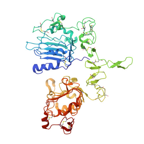
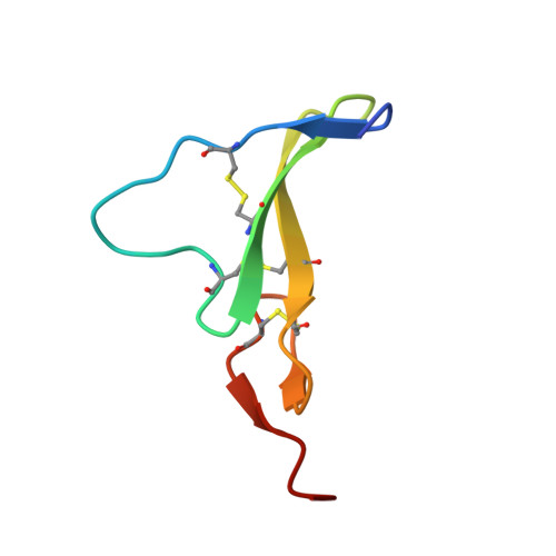
Crystal Structure of a Truncated Epidermal Growth Factor Receptor Extracellular Domain Bound to Transforming Growth Factor alpha

Garrett, T.P.J.McKern, N.M.Lou, M.Elleman, T.C.Adams, T.E.Lovrecz, G.O.Zhu, H.-J.Walker, F.Frenkel, M.J.Hoyne, P.A.Jorissen, R.N.Nice, E.C.Burgess, A.W.Ward, C.W.

(2002) Cell 110: 763-773

  • DOI: https://doi.org/10.1016/s0092-8674(02)00940-6
  • Primary Citation of Related Structures:  
    1MOX

  • PubMed Abstract: 

    We report the crystal structure, at 2.5 A resolution, of a truncated human EGFR ectodomain bound to TGFalpha. TGFalpha interacts with both L1 and L2 domains of EGFR, making many main chain contacts with L1 and interacting with L2 via key conserved residues. The results indicate how EGFR family members can bind a family of highly variable ligands. In the 2:2 TGFalpha:sEGFR501 complex, each ligand interacts with only one receptor molecule. There are two types of dimers in the asymmetric unit: a head-to-head dimer involving contacts between the L1 and L2 domains and a back-to-back dimer dominated by interactions between the CR1 domains of each receptor. Based on sequence conservation, buried surface area, and mutagenesis experiments, the back-to-back dimer is favored to be biologically relevant.


  • Organizational Affiliation

    Walter and Eliza Hall Institute of Medical Research, Post Office Royal Melbourne Hospital, Parkville, 3050, Victoria, Australia. tgarrett@wehi.edu.au


Macromolecules
Find similar proteins by:  (by identity cutoff)  |  3D Structure
Entity ID: 1
MoleculeChains Sequence LengthOrganismDetailsImage
Epidermal Growth Factor Receptor
A, B
501Homo sapiensMutation(s): 0 
Gene Names: EGFR
EC: 2.7.1.112
UniProt & NIH Common Fund Data Resources
Find proteins for P00533 (Homo sapiens)
Explore P00533 
Go to UniProtKB:  P00533
PHAROS:  P00533
GTEx:  ENSG00000146648 
Entity Groups  
Sequence Clusters30% Identity50% Identity70% Identity90% Identity95% Identity100% Identity
UniProt GroupP00533
Sequence Annotations
Expand
  • Reference Sequence
Find similar proteins by:  (by identity cutoff)  |  3D Structure
Entity ID: 2
MoleculeChains Sequence LengthOrganismDetailsImage
Transforming Growth Factor alpha
C, D
50Homo sapiensMutation(s): 0 
Gene Names: TGFA
UniProt & NIH Common Fund Data Resources
Find proteins for P01135 (Homo sapiens)
Explore P01135 
Go to UniProtKB:  P01135
PHAROS:  P01135
GTEx:  ENSG00000163235 
Entity Groups  
Sequence Clusters30% Identity50% Identity70% Identity90% Identity95% Identity100% Identity
UniProt GroupP01135
Sequence Annotations
Expand
  • Reference Sequence
Oligosaccharides

Help

Entity ID: 3
MoleculeChains Length2D Diagram Glycosylation3D Interactions
2-acetamido-2-deoxy-beta-D-glucopyranose-(1-4)-[alpha-L-fucopyranose-(1-6)]2-acetamido-2-deoxy-beta-D-glucopyranose
E
3N-Glycosylation
Glycosylation Resources
GlyTouCan:  G21290RB
GlyCosmos:  G21290RB
GlyGen:  G21290RB
Entity ID: 4
MoleculeChains Length2D Diagram Glycosylation3D Interactions
alpha-D-mannopyranose-(1-3)-beta-D-mannopyranose-(1-4)-2-acetamido-2-deoxy-beta-D-glucopyranose-(1-4)-2-acetamido-2-deoxy-beta-D-glucopyranose
F
4N-Glycosylation
Glycosylation Resources
GlyTouCan:  G81315DD
GlyCosmos:  G81315DD
GlyGen:  G81315DD
Entity ID: 5
MoleculeChains Length2D Diagram Glycosylation3D Interactions
beta-D-mannopyranose-(1-4)-2-acetamido-2-deoxy-beta-D-glucopyranose-(1-4)-[alpha-L-fucopyranose-(1-6)]2-acetamido-2-deoxy-beta-D-glucopyranose
G
4N-Glycosylation
Glycosylation Resources
GlyTouCan:  G32152BH
GlyCosmos:  G32152BH
GlyGen:  G32152BH
Entity ID: 6
MoleculeChains Length2D Diagram Glycosylation3D Interactions
2-acetamido-2-deoxy-beta-D-glucopyranose-(1-4)-2-acetamido-2-deoxy-beta-D-glucopyranose
H
2N-Glycosylation
Glycosylation Resources
GlyTouCan:  G42666HT
GlyCosmos:  G42666HT
GlyGen:  G42666HT
Small Molecules
Ligands 4 Unique
IDChains Name / Formula / InChI Key2D Diagram3D Interactions
NAG
Query on NAG

Download Ideal Coordinates CCD File 
S [auth B]2-acetamido-2-deoxy-beta-D-glucopyranose
C8 H15 N O6
OVRNDRQMDRJTHS-FMDGEEDCSA-N
PT
Query on PT

Download Ideal Coordinates CCD File 
I [auth A]
J [auth A]
K [auth A]
T [auth B]
U [auth B]
I [auth A],
J [auth A],
K [auth A],
T [auth B],
U [auth B],
V [auth B],
W [auth B]
PLATINUM (II) ION
Pt
HRGDZIGMBDGFTC-UHFFFAOYSA-N
CD
Query on CD

Download Ideal Coordinates CCD File 
AA [auth B]
BA [auth B]
DA [auth C]
EA [auth D]
L [auth A]
AA [auth B],
BA [auth B],
DA [auth C],
EA [auth D],
L [auth A],
M [auth A],
N [auth A],
O [auth A],
X [auth B],
Y [auth B],
Z [auth B]
CADMIUM ION
Cd
WLZRMCYVCSSEQC-UHFFFAOYSA-N
CL
Query on CL

Download Ideal Coordinates CCD File 
CA [auth B],
P [auth A],
Q [auth A],
R [auth A]
CHLORIDE ION
Cl
VEXZGXHMUGYJMC-UHFFFAOYSA-M
Experimental Data & Validation

Experimental Data

  • Method: X-RAY DIFFRACTION
  • Resolution: 2.50 Å
  • R-Value Free: 0.289 
  • R-Value Work: 0.237 
  • R-Value Observed: 0.240 
  • Space Group: P 1 21 1
Unit Cell:
Length ( Å )Angle ( ˚ )
a = 51.59α = 90
b = 198.71β = 102.03
c = 78.9γ = 90
Software Package:
Software NamePurpose
DENZOdata reduction
SCALEPACKdata scaling
MLPHAREphasing
CNSrefinement

Structure Validation

View Full Validation Report


Entry History 

Revision History  (Full details and data files)

  • Version 1.0: 2003-09-10
    Type: Initial release
  • Version 1.1: 2008-05-05
    Changes: Version format compliance
  • Version 1.2: 2011-07-13
    Changes: Non-polymer description, Version format compliance
  • Version 2.0: 2020-07-29
    Type: Remediation
    Reason: Carbohydrate remediation
    Changes: Atomic model, Data collection, Derived calculations, Structure summary