1MKV | pdb_00001mkv

CARBOXYLIC ESTER HYDROLASE COMPLEX (PLA2 + TRANSITION STATE ANALOG COMPLEX)


Experimental Data Snapshot

  • Method: X-RAY DIFFRACTION
  • Resolution: 1.89 Å
  • R-Value Work: 
    0.180 (Depositor), 0.191 (DCC) 
  • R-Value Observed: 
    0.180 (Depositor) 

Starting Model: experimental
View more details

wwPDB Validation 3D Report Full Report

Validation slider image for 1MKV

Ligand Structure Quality Assessment 


This is version 1.5 of the entry. See complete history

Literature

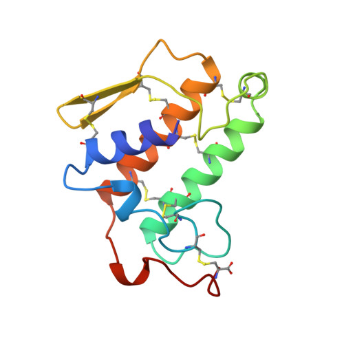
Structure of the complex of bovine pancreatic phospholipase A2 with a transition-state analogue.

Sekar, K.Kumar, A.Liu, X.Tsai, M.D.Gelb, M.H.Sundaralingam, M.

(1998) Acta Crystallogr D Biol Crystallogr 54: 334-341

  • DOI: https://doi.org/10.1107/s090744499701247x
  • Primary Citation Related Structures: 
    1MKV

  • PubMed Abstract: 

    The 1.89 A resolution structure of the complex of bovine pancreatic phospholipase A2 (PLA2) with the transition-state analogue L-1-O-octyl-2-heptylphosphonyl-sn-glycero-3-phosphoethanolamine (TSA) has been determined. The crystal of the complex is trigonal, space group P3121, a = b = 46.58 and c = 102.91 A and isomorphous to the native recombinant wild type (WT). The structure was refined to a final crystallographic R value of 18.0% including 957 protein atoms, 88 water molecules, one calcium ion and all 31 non-H atoms of the inhibitor at 1.89 A resolution. In all, 7 726 reflections [F>2sigma(F)] were used between 8.0 and 1.89 A resolution. The inhibitor is deeply locked into the active-site cleft and coordinates to the calcium ion by displacing the two water molecules in the calcium pentagonal bipyramid by the anionic O atoms of the phosphate and phosphonate group. The hydroxyl group of Tyr69 hydrogen bonds to the second anionic O atom of the phosphate group while that of the phosphonate group replaces the third water, 'catalytic' water, which forms a hydrogen bond to Ndelta1 of His48. The fourth water which also shares Ndelta1 of His48 is displaced by the steric hinderance of the inhibitor. The fifth conserved structural water is still present in the active site and forms a network of hydrogen bonds with the surrounding residues. The structure is compared to the other known TSA-PLA2 complexes.


  • Organizational Affiliation
    • Biological Macromolecular Structure Center, Departments of Chemistry and Biochemistry, 100 West 18th Avenue, The Ohio State University, Columbus, OH 43210-1185, USA.

Macromolecule Content 

  • Total Structure Weight: 14.34 kDa 
  • Atom Count: 1,077 
  • Modeled Residue Count: 123 
  • Deposited Residue Count: 123 
  • Unique protein chains: 1

Macromolecules

Find similar proteins by:|  3D Structure
Entity ID: 1
MoleculeChains  Sequence LengthOrganismDetailsImage
PHOSPHOLIPASE A2123Bos taurusMutation(s): 0 
Gene Names: MATURE PLA2
EC: 3.1.1.4
UniProt
Find proteins for P00593 (Bos taurus)
Explore P00593 
Go to UniProtKB:  P00593
Entity Groups
Sequence Clusters30% Identity50% Identity70% Identity90% Identity95% Identity100% Identity
UniProt GroupP00593
Sequence Annotations
Expand
Reference Sequence

Small Molecules

Ligands 2 Unique
IDChains Name / Formula / InChI Key2D Diagram3D Interactions
GEL

Query on GEL


Download:Ideal Coordinates CCD File
C [auth A]1-O-OCTYL-2-HEPTYLPHOSPHONYL-SN-GLYCERO-3-PHOSPHOETHANOLAMINE
C20 H45 N O8 P2
RCCNUBYROFOKAU-HXUWFJFHSA-N
CA

Query on CA


Download:Ideal Coordinates CCD File
B [auth A]CALCIUM ION
Ca
BHPQYMZQTOCNFJ-UHFFFAOYSA-N

Experimental Data & Validation

Experimental Data

  • Method: X-RAY DIFFRACTION
  • Resolution: 1.89 Å
  • R-Value Work:  0.180 (Depositor), 0.191 (DCC) 
  • R-Value Observed: 0.180 (Depositor) 
Space Group: P 31 2 1
Unit Cell:
Length ( Å )Angle ( ˚ )
a = 46.58α = 90
b = 46.58β = 90
c = 102.91γ = 120
Software Package:
Software NamePurpose
XENGENdata collection
XENGENdata reduction
X-PLORmodel building
X-PLORrefinement
XENGENdata scaling
X-PLORphasing

Structure Validation

View Full Validation Report


Ligand Structure Quality Assessment 


Entry History 

Deposition Data

Revision History  (Full details and data files)

  • Version 1.0: 1998-03-18
    Type: Initial release
  • Version 1.1: 2008-03-24
    Changes: Version format compliance
  • Version 1.2: 2011-07-13
    Changes: Version format compliance
  • Version 1.3: 2018-03-07
    Changes: Data collection, Other
  • Version 1.4: 2024-04-03
    Changes: Data collection, Database references, Derived calculations, Refinement description
  • Version 1.5: 2024-11-06
    Changes: Structure summary