1MAR | pdb_00001mar

REFINED 1.8 ANGSTROMS STRUCTURE OF HUMAN ALDOSE REDUCTASE COMPLEXED WITH THE POTENT INHIBITOR ZOPOLRESTAT


Experimental Data Snapshot

  • Method: X-RAY DIFFRACTION
  • Resolution: 1.80 Å
  • R-Value Work: 
    0.180 (Depositor) 
  • R-Value Observed: 
    0.180 (Depositor) 

wwPDB Validation 3D Report Full Report

Validation slider image for 1MAR

This is version 1.3 of the entry. See complete history

Literature

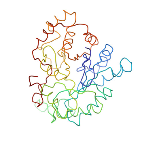
Refined 1.8 A structure of human aldose reductase complexed with the potent inhibitor zopolrestat.

Wilson, D.K.Tarle, I.Petrash, J.M.Quiocho, F.A.

(1993) Proc Natl Acad Sci U S A 90: 9847-9851

  • DOI: https://doi.org/10.1073/pnas.90.21.9847
  • Primary Citation Related Structures: 
    1MAR

  • PubMed Abstract: 

    As the action of aldose reductase (EC 1.1.1.21) is believed to be linked to the pathogenesis of diabetic complications affecting the nervous, renal, and visual systems, the development of therapeutic agents has attracted intense effort. We report the refined 1.8 A x-ray structure of the human holoenzyme complexed with zopolrestat, one of the most potent noncompetitive inhibitors. The zopolrestat fits snugly in the hydrophobic active site pocket and induces a hinge-flap motion of two peptide segments that closes the pocket. Excellent complementarity and affinity are achieved on inhibitor binding by the formation of 110 contacts (< or = 4 A) with 15 residues (10 hydrophobic), 13 with the NADPH coenzyme and 9 with four water molecules. The structure is key to understanding the mode of action of this class of inhibitors and for rational design of better therapeutics.


  • Organizational Affiliation
    • Howard Hughes Medical Institute, Baylor College of Medicine, Houston, TX 77030.

Macromolecule Content 

  • Total Structure Weight: 36.93 kDa 
  • Atom Count: 392 
  • Modeled Residue Count: 315 
  • Deposited Residue Count: 315 
  • Unique protein chains: 1

Macromolecules

Find similar proteins by:|  3D Structure
Entity ID: 1
MoleculeChains  Sequence LengthOrganismDetailsImage
ALDOSE REDUCTASE315Homo sapiensMutation(s): 0 
EC: 1.1.1.21 (PDB Primary Data), 1.1.1.372 (UniProt), 1.1.1.300 (UniProt), 1.1.1.54 (UniProt)
UniProt & NIH Common Fund Data Resources
Find proteins for P15121 (Homo sapiens)
Explore P15121 
Go to UniProtKB:  P15121
PHAROS:  P15121
GTEx:  ENSG00000085662 
Entity Groups
Sequence Clusters30% Identity50% Identity70% Identity90% Identity95% Identity100% Identity
UniProt GroupP15121
Sequence Annotations
Expand
Reference Sequence

Small Molecules

Ligands 2 Unique
IDChains Name / Formula / InChI Key2D Diagram3D Interactions
NAP

Query on NAP


Download:Ideal Coordinates CCD File
B [auth A]NADP NICOTINAMIDE-ADENINE-DINUCLEOTIDE PHOSPHATE
C21 H28 N7 O17 P3
XJLXINKUBYWONI-NNYOXOHSSA-N
ZST

Query on ZST


Download:Ideal Coordinates CCD File
C [auth A]3,4-DIHYDRO-4-OXO-3-((5-TRIFLUOROMETHYL-2-BENZOTHIAZOLYL)METHYL)-1-PHTHALAZINE ACETIC ACID
C19 H12 F3 N3 O3 S
BCSVCWVQNOXFGL-UHFFFAOYSA-N
Binding Affinity Annotations 
IDSourceBinding Affinity
ZST BindingDB:  1MAR Ki: 19 (nM) from 1 assay(s)
IC50: min: 1.9, max: 60 (nM) from 17 assay(s)

Experimental Data & Validation

Experimental Data

  • Method: X-RAY DIFFRACTION
  • Resolution: 1.80 Å
  • R-Value Work:  0.180 (Depositor) 
  • R-Value Observed: 0.180 (Depositor) 
Space Group: P 1
Unit Cell:
Length ( Å )Angle ( ˚ )
a = 47.64α = 64.47
b = 48.04β = 76.77
c = 40.48γ = 76.07
Software Package:
Software NamePurpose
X-PLORmodel building
X-PLORrefinement
X-PLORphasing

Structure Validation

View Full Validation Report


Entry History 

Deposition Data

Revision History  (Full details and data files)

  • Version 1.0: 1995-07-20
    Type: Initial release
  • Version 1.1: 2008-03-24
    Changes: Version format compliance
  • Version 1.2: 2011-07-13
    Changes: Version format compliance
  • Version 1.3: 2024-02-14
    Changes: Data collection, Database references, Derived calculations, Other