1LZY

X-RAY STRUCTURE OF TURKEY EGG LYSOZYME COMPLEX WITH DI-N-ACETYLCHITOBIOSE. RECOGNITION AND BINDING OF ALPHA-ANOMERIC FORM


Experimental Data Snapshot

  • Method: X-RAY DIFFRACTION
  • Resolution: 1.55 Å
  • R-Value Work: 0.175 
  • R-Value Observed: 0.175 

wwPDB Validation   3D Report Full Report


This is version 2.0 of the entry. See complete history


Literature

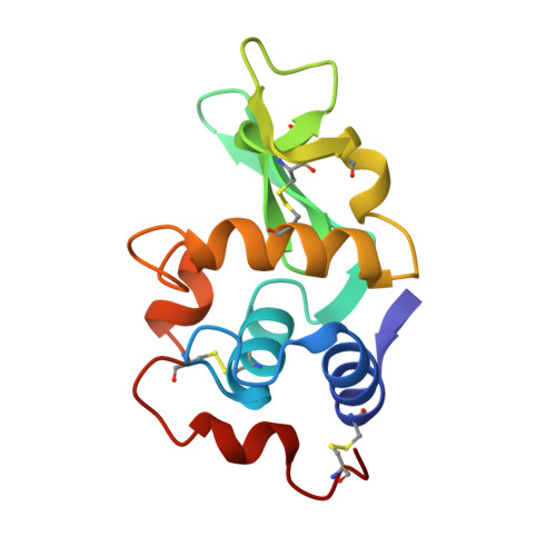
X-ray structure of monoclinic turkey egg lysozyme at 1.3 A resolution.

Harata, K.

(1993) Acta Crystallogr D Biol Crystallogr 49: 497-504

  • DOI: https://doi.org/10.1107/S0907444993005542
  • Primary Citation of Related Structures:  
    135L, 1LZY

  • PubMed Abstract: 

    Monoclinic crystals of turkey egg lysozyme (TEL, E.C. 3.2.1.17) were obtained from 2.2 M ammonium sulfate solution at pH 4.2. They belong to space group P2(1) with unit-cell dimensions a = 38.07, b = 33.20, c = 46.12 A and beta = 110.1 degrees, and contain one molecule in the asymmetric unit (V(m) = 1.91 A(3) Da(-1)). The three-dimensional structure of TEL was solved by the method of multiple isomorphous replacement with anomalous scattering. Area detector data to 1.5 A resolution from native and heavy-atom derivatives were used for the structure determination. The structure was refined by the simulated-annealing method with diffraction data of 10-1.30 A resolution. The conventional R factor was 0.189. The root-mean-square deviations from ideal bond distances and angles were 0.016 A and 2.9 degrees, respectively. The backbone structure of TEL is very similar to that of hen egg lysozyme (HEL) and the difference in seven amino-acid residues does not affect the basic folding of the polypeptide chain. Except for the region from Gly101 to Gly104, the geometry of the active-site cleft is conserved between TEL and HEL. The Gly101 residue is located at the end of the sugar-binding site and the structural change in this region between TEL and HEL is considered to be responsible for the difference in their enzymatic properties.


  • Organizational Affiliation

    Biomolecules Department, National Institute of Bioscience and Human Technology, Tsukuba, Ibaraki, Japan.


Macromolecules
Find similar proteins by:  (by identity cutoff)  |  3D Structure
Entity ID: 1
MoleculeChains Sequence LengthOrganismDetailsImage
TURKEY EGG WHITE LYSOZYME129Meleagris gallopavoMutation(s): 0 
EC: 3.2.1.17
UniProt
Find proteins for P00703 (Meleagris gallopavo)
Explore P00703 
Go to UniProtKB:  P00703
Entity Groups  
Sequence Clusters30% Identity50% Identity70% Identity90% Identity95% Identity100% Identity
UniProt GroupP00703
Sequence Annotations
Expand
  • Reference Sequence
Oligosaccharides

Help

Entity ID: 2
MoleculeChains Length2D Diagram Glycosylation3D Interactions
2-acetamido-2-deoxy-beta-D-glucopyranose-(1-4)-2-acetamido-2-deoxy-alpha-D-glucopyranose
B
2N/A
Glycosylation Resources
GlyTouCan:  G05026ZL
GlyCosmos:  G05026ZL
GlyGen:  G05026ZL
Experimental Data & Validation

Experimental Data

  • Method: X-RAY DIFFRACTION
  • Resolution: 1.55 Å
  • R-Value Work: 0.175 
  • R-Value Observed: 0.175 
  • Space Group: P 1 21 1
Unit Cell:
Length ( Å )Angle ( ˚ )
a = 38.22α = 90
b = 33.14β = 109.9
c = 46γ = 90
Software Package:
Software NamePurpose
X-PLORmodel building
X-PLORrefinement
X-PLORphasing

Structure Validation

View Full Validation Report


Entry History 

Deposition Data

  • Released Date: 1995-02-27 
  • Deposition Author(s): Harata, K.

Revision History  (Full details and data files)

  • Version 1.0: 1995-02-27
    Type: Initial release
  • Version 1.1: 2008-03-24
    Changes: Version format compliance
  • Version 1.2: 2011-07-13
    Changes: Non-polymer description, Version format compliance
  • Version 1.3: 2017-11-29
    Changes: Derived calculations, Other
  • Version 2.0: 2020-07-29
    Type: Remediation
    Reason: Carbohydrate remediation
    Changes: Atomic model, Data collection, Derived calculations, Structure summary