Structural studies of metarhodopsin II, the activated form of the G-protein coupled receptor, rhodopsin.
Choi, G., Landin, J., Galan, J.F., Birge, R.R., Albert, A.D., Yeagle, P.L.(2002) Biochemistry 41: 7318-7324
- PubMed: 12044163 Search on PubMed
- DOI: https://doi.org/10.1021/bi025507w
- Primary Citation Related Structures:
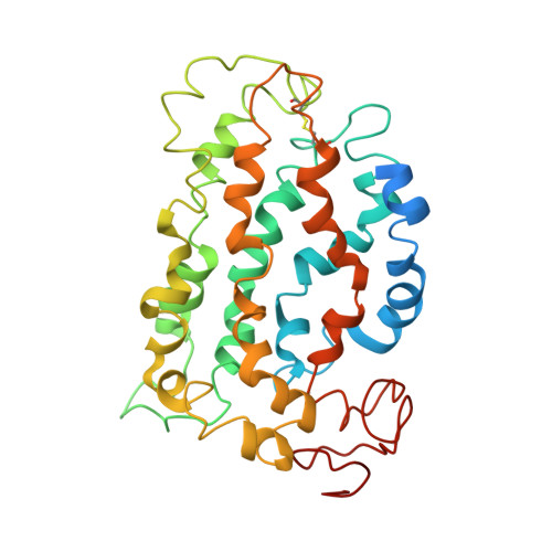
1LN6 - PubMed Abstract:
The structural changes that accompany activation of a G-protein coupled receptor (GPCR) are not well understood. To better understand the activation of rhodopsin, the GPCR responsible for visual transduction, we report studies on the three-dimensional structure for the activated state of this receptor, metarhodopsin II. Differences between the three-dimensional structure of ground state rhodopsin and metarhodopsin II, particularly in the cytoplasmic face of the receptor, suggest how the receptor is activated to couple with transducin. In particular, activation opens a groove on the surface of the receptor that could bind the N-terminal helix of the G protein, transducin alpha.
- Department of Molecular and Cell Biology, University of Connecticut, Storrs, CT 06269, USA.
Organizational Affiliation: