Crystallographic dissection of the thermal motion of protein-sugar complex.
Harata, K., Kanai, R.(2002) Proteins 48: 53-62
- PubMed: 12012337 Search on PubMed
- DOI: https://doi.org/10.1002/prot.10127
- Primary Citation Related Structures:
1LJN - PubMed Abstract:
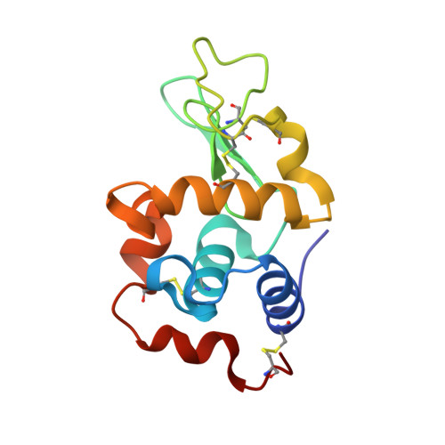
The crystal structure of turkey egg lysozyme (TEL) complexed with di-N-acetylchitobiose (NAG2) was refined at 1.19 A resolution by the full-matrix least-squares method with anisotropic temperature factors, and its thermal motion was evaluated by the TLS method. The average ESDs of atomic parameters of nonhydrogen atoms were 0.030 A for coordinates and 0.025 A(2) for anisotropic temperature factors. The active site cleft of TEL binds the alpha-anomer of NAG2 in a nonproductive binding mode with its pyranose rings parallel to a beta-sheet. The TEL structure was compared with the re-refined 1.12 A structure of native TEL. The RMS difference for equivalent Calpha atoms was 0.103 A and a relatively large difference was observed in the region of residues 104-125 rather than in the beta-sheet region where NAG2 was bound. In contrast, the temperature factor of the beta-sheet region was significantly decreased by the NAG2 binding. The TLS model that describes the rigid body motion in translation, libration, and screw motion was adopted for the evaluation of the molecular motion of TEL and NAG2, and the TLS parameters were determined by the least-squares fit to U(ij). The contribution of the external motion of TEL was estimated to be 55.8% of the observed temperature factor for the native structure and 45.9% for the NAG2 complex. The internal motion of TEL represented with atomic thermal ellipsoids was very similar between the native and complex structures except the NAG2 binding region. In the structure of NAG2, the rigid body motion dominates the thermal motion. The center of rotation of NAG2, 4.45A far from the center of gravity, is on the nitrogen atom of the acetylamino group that is hydrogen bonded to the main-chain peptide groups of Asn49 and Ala107. The rigid body motion of NAG2 indicates that the acetylamino group is most strongly bound to the active site, and the recognition of this group is a crucial step of the substrate binding.
- Biological Information Research Center, National Institute of Advanced Industrial Science and Technology, Tsukuba, Ibaraki, Japan. k-harata@aist.go.jp
Organizational Affiliation: