1LAJ

The Structure of Tomato Aspermy Virus by X-Ray Crystallography


Experimental Data Snapshot

  • Method: X-RAY DIFFRACTION
  • Resolution: 3.40 Å
  • R-Value Free: 0.228 
  • R-Value Work: 0.218 

wwPDB Validation   3D Report Full Report


This is version 1.3 of the entry. See complete history


Literature

The Structure of Tomato Aspermy Virus by X-Ray Crystallography

Lucas, R.W.Larson, S.B.Canady, M.A.McPherson, A.

(2002) J Struct Biol 139: 90-102

  • DOI: https://doi.org/10.1016/s1047-8477(02)00561-0
  • Primary Citation of Related Structures:  
    1LAJ

  • PubMed Abstract: 

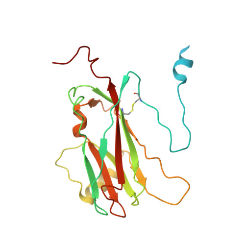
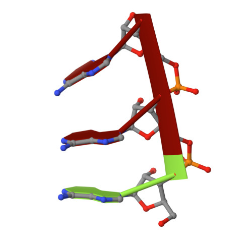
    The three-dimensional structure of tomato aspermy virus (TAV) has been solved by X-ray crystallography and refined to an R factor of 0.218 for 3.4-40 A data (effective resolution of 4A). Molecular replacement, using cucumber mosaic virus (Smith et al., 2000), provided phases for the initial maps used for model building. The coat protein of the 280 A diameter virion has the canonical "Swiss roll" beta-barrel topology with a distinctive amino-terminal alpha-helix directed into the interior of the virus where it interacts with encapsidated RNA. The N-terminal helices are joined to the beta-barrels of protein subunits by extended polypeptides of six amino acids, which serve as flexible hinges allowing movement of the helices in response to local RNA distribution. Segments of three nucleotides of partially disordered RNA interact with the capsid, primarily through arginine residues, at interfaces between A and B subunits. Side chains of cys64 and cys106 form the first disulfide observed in a cucumovirus, including a unique cysteine, 106, in a region otherwise conserved. A positive ion, putatively modeled as a Mg(+)ion, lies on the quasi-threefold axis surrounded by three quasi-symmetric glutamate 175 side chains.


  • Organizational Affiliation

    Department of Molecular Biology and Biochemistry, University of California, Irvine, Irvine, CA 92697-3900, USA.


Macromolecules

Find similar proteins by:  (by identity cutoff)  |  3D Structure
Entity ID: 2
MoleculeChains Sequence LengthOrganismDetailsImage
capsid proteinB [auth A],
C [auth B],
D [auth C]
217Tomato aspermy virusMutation(s): 0 
UniProt
Find proteins for P23627 (Tomato aspermy virus)
Explore P23627 
Go to UniProtKB:  P23627
Entity Groups  
Sequence Clusters30% Identity50% Identity70% Identity90% Identity95% Identity100% Identity
UniProt GroupP23627
Sequence Annotations
Expand
  • Reference Sequence

Find similar nucleic acids by:  Sequence   |   3D Structure  

Entity ID: 1
MoleculeChains LengthOrganismImage
5'-R(*AP*AP*A)-3'A [auth R]3N/A
Sequence Annotations
Expand
  • Reference Sequence
Experimental Data & Validation

Experimental Data

  • Method: X-RAY DIFFRACTION
  • Resolution: 3.40 Å
  • R-Value Free: 0.228 
  • R-Value Work: 0.218 
  • Space Group: I 2 2 2
Unit Cell:
Length ( Å )Angle ( ˚ )
a = 294.296α = 90
b = 327.393β = 90
c = 382.066γ = 90
Software Package:
Software NamePurpose
DENZOdata reduction
SCALEPACKdata scaling
X-PLORmodel building
X-PLORrefinement
X-PLORphasing

Structure Validation

View Full Validation Report


Entry History 

Deposition Data

Revision History  (Full details and data files)

  • Version 1.0: 2002-11-27
    Type: Initial release
  • Version 1.1: 2008-04-28
    Changes: Version format compliance
  • Version 1.2: 2011-07-13
    Changes: Version format compliance
  • Version 1.3: 2023-08-16
    Changes: Data collection, Database references, Derived calculations, Refinement description