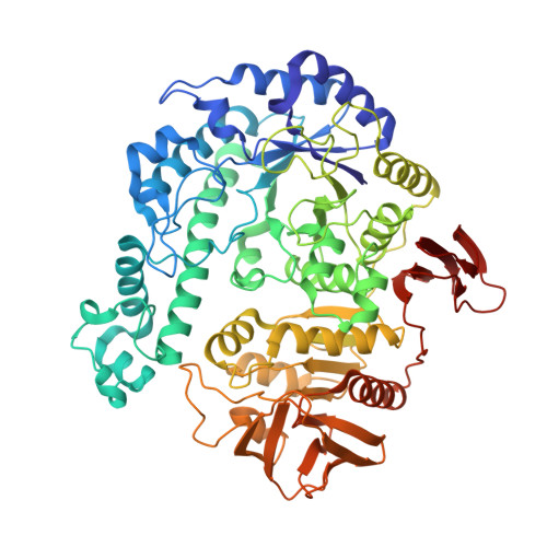
1KWG

Crystal structure of Thermus thermophilus A4 beta-galactosidase


Experimental Data Snapshot

  • Method: X-RAY DIFFRACTION
  • Resolution: 1.60 Å
  • R-Value Free: 0.184 
  • R-Value Work: 0.167 
  • R-Value Observed: 0.167 

wwPDB Validation   3D Report Full Report


This is version 1.2 of the entry. See complete history


Literature

Trimeric Crystal Structure of the Glycoside Hydrolase Family 42 beta-Galactosidase from Thermus thermophilus A4 and the Structure of its Complex with Galactose

Hidaka, M.Fushinobu, S.Ohtsu, N.Motoshima, H.Matsuzawa, H.Shoun, H.Wakagi, T.

(2002) J Mol Biol 322: 79-91

  • DOI: https://doi.org/10.1016/s0022-2836(02)00746-5
  • Primary Citation of Related Structures:  
    1KWG, 1KWK

  • PubMed Abstract: 
  • The beta-galactosidase from an extreme thermophile, Thermus thermophilus A4 (A4-beta-Gal), is thermostable and belongs to the glycoside hydrolase family 42 (GH-42). As the first known structures of a GH-42 enzyme, we determined the crystal structures of free and galactose-bound A4-beta-Gal at 1 ...

    The beta-galactosidase from an extreme thermophile, Thermus thermophilus A4 (A4-beta-Gal), is thermostable and belongs to the glycoside hydrolase family 42 (GH-42). As the first known structures of a GH-42 enzyme, we determined the crystal structures of free and galactose-bound A4-beta-Gal at 1.6A and 2.2A resolution, respectively. A4-beta-Gal forms a homotrimeric structure resembling a flowerpot. Each monomer has an active site located inside a large central tunnel. The N-terminal domain of A4-beta-Gal has a TIM barrel fold, as predicted from hydrophobic cluster analysis. The putative catalytic residues of A4-beta-Gal (Glu141 and Glu312) superimpose well with the catalytic residues of Escherichia coli beta-galactosidase. The environment around the catalytic nucleophile (Glu312) is similar to that in the case of E.coli beta-galactosidase, but the recognition mechanism for a substrate is different. Trp182 of the next subunit of the trimer constitutes a part of the active-site pocket, indicating that the trimeric structure is essential for the enzyme activity. Structural comparison with other glycoside hydrolases revealed that many features of the 4/7 superfamily are conserved in the A4-beta-Gal structure. On the basis of the results of 1H NMR spectroscopy, A4-beta-Gal was determined to be a "retaining" enzyme. Interestingly, the active site was similar with those of retaining enzymes, but the overall fold of the TIM barrel domain was very similar to that of an inverting enzyme, beta-amylase.


    Related Citations: 
    • Thermostable beta-galactosidase from an extreme thermophile, Thermus sp. A4: enzyme purification and characterization, and gene cloning and sequencing
      Ohtsu, N., Motoshima, H., Goto, K., Tsukasaki, F., Matsuzawa, H.
      (1998) Biosci Biotechnol Biochem 62: 1539

    Organizational Affiliation

    Department of Biotechnology, The University of Tokyo, Japan.


Macromolecules
Find similar proteins by:  (by identity cutoff)  |  3D Structure
Entity ID: 1
MoleculeChainsSequence LengthOrganismDetailsImage
BETA-GALACTOSIDASEA645Thermus thermophilusMutation(s): 0 
EC: 3.2.1.23
UniProt
Find proteins for O69315 (Thermus thermophilus)
Explore O69315 
Go to UniProtKB:  O69315
Entity Groups  
Sequence Clusters30% Identity50% Identity70% Identity90% Identity95% Identity100% Identity
UniProt GroupO69315
Protein Feature View
Expand
  • Reference Sequence
Small Molecules
Ligands 4 Unique
IDChainsName / Formula / InChI Key2D Diagram3D Interactions
MPD
Query on MPD

Download Ideal Coordinates CCD File 
F [auth A],
G [auth A]
(4S)-2-METHYL-2,4-PENTANEDIOL
C6 H14 O2
SVTBMSDMJJWYQN-YFKPBYRVSA-N
 Ligand Interaction
ZN
Query on ZN

Download Ideal Coordinates CCD File 
E [auth A]ZINC ION
Zn
PTFCDOFLOPIGGS-UHFFFAOYSA-N
 Ligand Interaction
ACT
Query on ACT

Download Ideal Coordinates CCD File 
C [auth A],
D [auth A]
ACETATE ION
C2 H3 O2
QTBSBXVTEAMEQO-UHFFFAOYSA-M
 Ligand Interaction
CL
Query on CL

Download Ideal Coordinates CCD File 
B [auth A]CHLORIDE ION
Cl
VEXZGXHMUGYJMC-UHFFFAOYSA-M
 Ligand Interaction
Experimental Data & Validation

Experimental Data

  • Method: X-RAY DIFFRACTION
  • Resolution: 1.60 Å
  • R-Value Free: 0.184 
  • R-Value Work: 0.167 
  • R-Value Observed: 0.167 
  • Space Group: P 3 2 1
Unit Cell:
Length ( Å )Angle ( ˚ )
a = 97.737α = 90
b = 97.737β = 90
c = 129.37γ = 120
Software Package:
Software NamePurpose
SOLVEphasing
CNSrefinement
HKL-2000data reduction
CCP4data scaling

Structure Validation

View Full Validation Report


Entry History 

Deposition Data

Revision History  (Full details and data files)

  • Version 1.0: 2002-09-18
    Type: Initial release
  • Version 1.1: 2008-04-27
    Changes: Version format compliance
  • Version 1.2: 2011-07-13
    Changes: Derived calculations, Version format compliance