1KVR | pdb_00001kvr

UDP-GALACTOSE 4-EPIMERASE COMPLEXED WITH UDP-PHENOL


Experimental Data Snapshot

  • Method: X-RAY DIFFRACTION
  • Resolution: 1.90 Å
  • R-Value Work: 
    0.179 (Depositor) 

wwPDB Validation 3D Report Full Report

Validation slider image for 1KVR

This is version 1.6 of the entry. See complete history

Literature

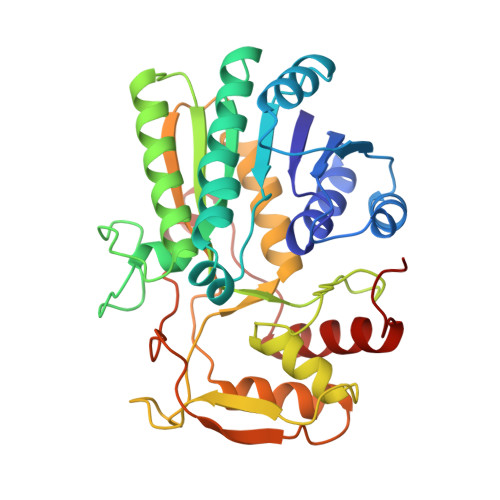
Molecular structures of the S124A, S124T, and S124V site-directed mutants of UDP-galactose 4-epimerase from Escherichia coli.

Thoden, J.B.Gulick, A.M.Holden, H.M.

(1997) Biochemistry 36: 10685-10695

  • DOI: https://doi.org/10.1021/bi9704313
  • Primary Citation Related Structures: 
    1KVQ, 1KVR, 1KVS, 1KVT

  • PubMed Abstract: 

    UDP-galactose 4-epimerase plays a critical role in sugar metabolism by catalyzing the interconversion of UDP-galactose and UDP-glucose. Originally, it was assumed that the enzyme contained a "traditional" catalytic base that served to abstract a proton from the 4'-hydroxyl group of the UDP-glucose or UDP-galactose substrates during the course of the reaction. However, recent high-resolution X-ray crystallographic analyses of the protein from Escherichia coli have demonstrated the lack of an aspartate, a glutamate, or a histidine residue properly oriented within the active site cleft for serving such a functional role. Rather, the X-ray crystallographic investigation of the epimerase.NADH.UDP-glucose abortive complex from this laboratory has shown that both Ser 124 and Tyr 149 are located within hydrogen bonding distance to the 4'- and 3'-hydroxyl groups of the sugar, respectively. To test the structural role of Ser 124 in the reaction mechanism of epimerase, three site-directed mutant proteins, namely S124A, S124T, and S124V, were constructed and crystals of the S124A.NADH.UDP, S124A.NADH.UDP-glucose, S124T. NADH.UDP-glucose, and S124V.NADH.UDP-glucose complexes were grown. All of the crystals employed in this investigation belonged to the space group P3221 with the following unit cell dimensions: a = b = 83.8 A, c = 108.4 A, and one subunit per asymmetric unit. X-ray data sets were collected to at least 2.15 A resolution, and each protein model was subsequently refined to an R value of lower than 19.0% for all measured X-ray data. The investigations described here demonstrate that the decreases in enzymatic activities observed for these mutant proteins are due to the loss of a properly positioned hydroxyl group at position 124 and not to major tertiary and quaternary structural perturbations. In addition, these structures demonstrate the importance of a hydroxyl group at position 124 in stabilizing the anti conformation of the nicotinamide ring as observed in the previous structural analysis of the epimerase.NADH. UDP complex.


  • Organizational Affiliation
    • Institute for Enzyme Research, Graduate School, University of Wisconsin, Madison 53705, USA.

Macromolecule Content 

  • Total Structure Weight: 38.62 kDa 
  • Atom Count: 3,241 
  • Modeled Residue Count: 338 
  • Deposited Residue Count: 338 
  • Unique protein chains: 1

Macromolecules

Find similar proteins by:|  3D Structure
Entity ID: 1
MoleculeChains  Sequence LengthOrganismDetailsImage
UDP-GALACTOSE 4-EPIMERASE338Escherichia coliMutation(s): 1 
EC: 5.1.3.2
UniProt
Find proteins for P09147 (Escherichia coli (strain K12))
Explore P09147 
Go to UniProtKB:  P09147
Entity Groups
Sequence Clusters30% Identity50% Identity70% Identity90% Identity95% Identity100% Identity
UniProt GroupP09147
Sequence Annotations
Expand
Reference Sequence

Small Molecules

Ligands 5 Unique
IDChains Name / Formula / InChI Key2D Diagram3D Interactions
NAD

Query on NAD


Download:Ideal Coordinates CCD File
F [auth A]NICOTINAMIDE-ADENINE-DINUCLEOTIDE
C21 H27 N7 O14 P2
BAWFJGJZGIEFAR-NNYOXOHSSA-N
UDP

Query on UDP


Download:Ideal Coordinates CCD File
G [auth A]URIDINE-5'-DIPHOSPHATE
C9 H14 N2 O12 P2
XCCTYIAWTASOJW-XVFCMESISA-N
PEG

Query on PEG


Download:Ideal Coordinates CCD File
I [auth A]DI(HYDROXYETHYL)ETHER
C4 H10 O3
MTHSVFCYNBDYFN-UHFFFAOYSA-N
EDO

Query on EDO


Download:Ideal Coordinates CCD File
H [auth A]1,2-ETHANEDIOL
C2 H6 O2
LYCAIKOWRPUZTN-UHFFFAOYSA-N
NA

Query on NA


Download:Ideal Coordinates CCD File
B [auth A],
C [auth A],
D [auth A],
E [auth A]
SODIUM ION
Na
FKNQFGJONOIPTF-UHFFFAOYSA-N

Experimental Data & Validation

Experimental Data

  • Method: X-RAY DIFFRACTION
  • Resolution: 1.90 Å
  • R-Value Work:  0.179 (Depositor) 
Space Group: P 32 2 1
Unit Cell:
Length ( Å )Angle ( ˚ )
a = 83.5α = 90
b = 83.5β = 90
c = 108.4γ = 120
Software Package:
Software NamePurpose
TNTrefinement
XSCALIBREdata scaling

Structure Validation

View Full Validation Report


Entry History 

Deposition Data

Revision History  (Full details and data files)

  • Version 1.0: 1998-03-18
    Type: Initial release
  • Version 1.1: 2008-03-03
    Changes: Version format compliance
  • Version 1.2: 2011-07-13
    Changes: Derived calculations, Version format compliance
  • Version 1.3: 2011-11-16
    Changes: Atomic model
  • Version 1.4: 2015-05-27
    Changes: Structure summary
  • Version 1.5: 2021-11-03
    Changes: Database references, Derived calculations, Other
  • Version 1.6: 2024-02-14
    Changes: Data collection