1KTK | pdb_00001ktk

Complex of Streptococcal pyrogenic enterotoxin C (SpeC) with a human T cell receptor beta chain (Vbeta2.1)


Experimental Data Snapshot

  • Method: X-RAY DIFFRACTION
  • Resolution: 3.00 Å
  • R-Value Free: 
    0.334 (Depositor), 0.341 (DCC) 
  • R-Value Work: 
    0.326 (Depositor), 0.334 (DCC) 
  • R-Value Observed: 
    0.326 (Depositor) 

wwPDB Validation 3D Report Full Report

Validation slider image for 1KTK

This is version 1.3 of the entry. See complete history

Literature

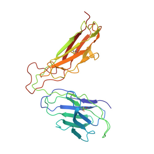
Structures of two streptococcal superantigens bound to TCR beta chains reveal diversity in the architecture of T cell signaling complexes.

Sundberg, E.J.Li, H.Llera, A.S.McCormick, J.K.Tormo, J.Schlievert, P.M.Karjalainen, K.Mariuzza, R.A.

(2002) Structure 10: 687-699

  • DOI: https://doi.org/10.1016/s0969-2126(02)00759-1
  • Primary Citation Related Structures: 
    1KTK, 1L0X, 1L0Y

  • PubMed Abstract: 

    Superantigens (SAGs) crosslink MHC class II and TCR molecules, resulting in an overstimulation of T cells associated with human disease. SAGs interact with several different surfaces on MHC molecules, necessitating the formation of multiple distinct MHC-SAG-TCR ternary signaling complexes. Variability in SAG-TCR binding modes could also contribute to the structural heterogeneity of SAG-dependent signaling complexes. We report crystal structures of the streptococcal SAGs SpeA and SpeC in complex with their corresponding TCR beta chain ligands that reveal distinct TCR binding modes. The SpeC-TCR beta chain complex structure, coupled with the recently determined SpeC-HLA-DR2a complex structure, provides a model for a novel T cell signaling complex that precludes direct TCR-MHC interactions. Thus, highly efficient T cell activation may be achieved through structurally diverse strategies of TCR ligation.


  • Organizational Affiliation
    • Center for Advanced Research in Biotechnology, W.M. Keck Laboratory for Structural Biology, University of Maryland Biotechnology Institute, 9600 Gudelsky Drive, Rockville, MD 20850, USA.

Macromolecule Content 

  • Total Structure Weight: 152.46 kDa 
  • Atom Count: 9,511 
  • Modeled Residue Count: 1,200 
  • Deposited Residue Count: 1,326 
  • Unique protein chains: 2

Macromolecules

Find similar proteins by:|  3D Structure
Entity ID: 1
MoleculeChains  Sequence LengthOrganismDetailsImage
Exotoxin type C
A, B, C, D
208Streptococcus pyogenesMutation(s): 0 
UniProt
Find proteins for Q8NKX2 (Streptococcus pyogenes serotype M18 (strain MGAS8232))
Explore Q8NKX2 
Go to UniProtKB:  Q8NKX2
Entity Groups
Sequence Clusters30% Identity50% Identity70% Identity90% Identity95% Identity100% Identity
UniProt GroupQ8NKX2
Sequence Annotations
Expand
Reference Sequence
Find similar proteins by:|  3D Structure
Entity ID: 2
MoleculeChains  Sequence LengthOrganismDetailsImage
T-cell receptor beta chain
E, F
247Homo sapiensMutation(s): 0 
UniProt
Find proteins for P01850 (Homo sapiens)
Explore P01850 
Go to UniProtKB:  P01850
Entity Groups
Sequence Clusters30% Identity50% Identity70% Identity90% Identity95% Identity100% Identity
UniProt GroupP01850
Sequence Annotations
Expand
Reference Sequence

Experimental Data & Validation

Experimental Data

  • Method: X-RAY DIFFRACTION
  • Resolution: 3.00 Å
  • R-Value Free:  0.334 (Depositor), 0.341 (DCC) 
  • R-Value Work:  0.326 (Depositor), 0.334 (DCC) 
  • R-Value Observed: 0.326 (Depositor) 
Space Group: P 1 21 1
Unit Cell:
Length ( Å )Angle ( ˚ )
a = 57.378α = 90
b = 146.764β = 98.61
c = 135.69γ = 90
Software Package:
Software NamePurpose
DENZOdata reduction
SCALEPACKdata scaling
AMoREphasing
CNSrefinement

Structure Validation

View Full Validation Report


Entry History 

Deposition Data

Revision History  (Full details and data files)

  • Version 1.0: 2002-06-07
    Type: Initial release
  • Version 1.1: 2008-04-27
    Changes: Version format compliance
  • Version 1.2: 2011-07-13
    Changes: Version format compliance
  • Version 1.3: 2024-11-20
    Changes: Data collection, Database references, Structure summary