Structure and function of threonine synthase from yeast.
Garrido-Franco, M., Ehlert, S., Messerschmidt, A., Marinkovic, S., Huber, R., Laber, B., Bourenkov, G.P., Clausen, T.(2002) J Biol Chem 277: 12396-12405
- PubMed: 11756443 Search on PubMed
- DOI: https://doi.org/10.1074/jbc.M108734200
- Primary Citation Related Structures:
1KL7 - PubMed Abstract:
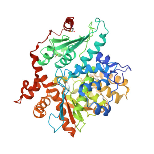
Threonine synthase catalyzes the final step of threonine biosynthesis, the pyridoxal 5'-phosphate (PLP)-dependent conversion of O-phosphohomoserine into threonine and inorganic phosphate. Threonine is an essential nutrient for mammals, and its biosynthetic machinery is restricted to bacteria, plants, and fungi; therefore, threonine synthase represents an interesting pharmaceutical target. The crystal structure of threonine synthase from Saccharomyces cerevisiae has been solved at 2.7 A resolution using multiwavelength anomalous diffraction. The structure reveals a monomer as active unit, which is subdivided into three distinct domains: a small N-terminal domain, a PLP-binding domain that covalently anchors the cofactor and a so-called large domain, which contains the main of the protein body. All three domains show the typical open alpha/beta architecture. The cofactor is bound at the interface of all three domains, buried deeply within a wide canyon that penetrates the whole molecule. Based on structural alignments with related enzymes, an enzyme-substrate complex was modeled into the active site of yeast threonine synthase, which revealed essentials for substrate binding and catalysis. Furthermore, the comparison with related enzymes of the beta-family of PLP-dependent enzymes indicated structural determinants of the oligomeric state and thus rationalized for the first time how a PLP enzyme acts in monomeric form.
- Max-Planck-Institut für Biochemie, Abteilung Strukturforschung, am Klopferspitz 18A, Martinsried 82152, Germany.
Organizational Affiliation: