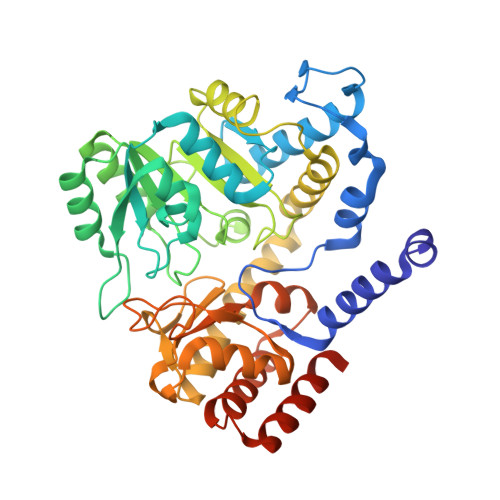
Crystal structure of binary and ternary complexes of serine hydroxymethyltransferase from Bacillus stearothermophilus: insights into the catalytic mechanism.
Trivedi, V., Gupta, A., Jala, V.R., Saravanan, P., Rao, G.S., Rao, N.A., Savithri, H.S., Subramanya, H.S.(2002) J Biological Chem 277: 17161-17169
- PubMed: 11877399
- DOI: https://doi.org/10.1074/jbc.M111976200
- Primary Citation of Related Structures:
1KKJ, 1KKP, 1KL1, 1KL2 - PubMed Abstract:
Serine hydroxymethyltransferase (SHMT), a member of the alpha-class of pyridoxal phosphate-dependent enzymes, catalyzes the reversible conversion of serine to glycine and tetrahydrofolate to 5,10-methylene tetrahydrofolate. We present here the crystal structures of the native enzyme and its complexes with serine, glycine, glycine, and 5-formyl tetrahydrofolate (FTHF) from Bacillus stearothermophilus. The first structure of the serine-bound form of SHMT allows identification of residues involved in serine binding and catalysis. The SHMT-serine complex does not show any significant conformational change compared with the native enzyme, contrary to that expected for a conversion from an "open" to "closed" form of the enzyme. However, the ternary complex with FTHF and glycine shows the reported conformational changes. In contrast to the Escherichia coli enzyme, this complex shows asymmetric binding of the FTHF to the two monomers within the dimer in a way similar to the murine SHMT. Comparison of the ternary complex with the native enzyme reveals the structural basis for the conformational change and asymmetric binding of FTHF. The four structures presented here correspond to the various reaction intermediates of the catalytic pathway and provide evidence for a direct displacement mechanism for the hydroxymethyl transfer rather than a retroaldol cleavage.
- Molecular and Structural Biology Division, Central Drug Research Institute, Chattar Manzil Palace, Mahatma Gandhi Marg, P. B. No. 173, Lucknow 226001, India.
Organizational Affiliation:


