1KKP

Crystal Structure of Serine Hydroxymethyltransferase complexed with Serine


Experimental Data Snapshot

  • Method: X-RAY DIFFRACTION
  • Resolution: 1.93 Å
  • R-Value Free: 0.206 
  • R-Value Work: 0.179 

wwPDB Validation   3D Report Full Report


Ligand Structure Quality Assessment 


This is version 1.3 of the entry. See complete history


Literature

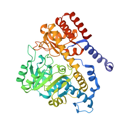
Crystal structure of binary and ternary complexes of serine hydroxymethyltransferase from Bacillus stearothermophilus: insights into the catalytic mechanism.

Trivedi, V.Gupta, A.Jala, V.R.Saravanan, P.Rao, G.S.Rao, N.A.Savithri, H.S.Subramanya, H.S.

(2002) J Biol Chem 277: 17161-17169

  • DOI: https://doi.org/10.1074/jbc.M111976200
  • Primary Citation of Related Structures:  
    1KKJ, 1KKP, 1KL1, 1KL2

  • PubMed Abstract: 

    Serine hydroxymethyltransferase (SHMT), a member of the alpha-class of pyridoxal phosphate-dependent enzymes, catalyzes the reversible conversion of serine to glycine and tetrahydrofolate to 5,10-methylene tetrahydrofolate. We present here the crystal structures of the native enzyme and its complexes with serine, glycine, glycine, and 5-formyl tetrahydrofolate (FTHF) from Bacillus stearothermophilus. The first structure of the serine-bound form of SHMT allows identification of residues involved in serine binding and catalysis. The SHMT-serine complex does not show any significant conformational change compared with the native enzyme, contrary to that expected for a conversion from an "open" to "closed" form of the enzyme. However, the ternary complex with FTHF and glycine shows the reported conformational changes. In contrast to the Escherichia coli enzyme, this complex shows asymmetric binding of the FTHF to the two monomers within the dimer in a way similar to the murine SHMT. Comparison of the ternary complex with the native enzyme reveals the structural basis for the conformational change and asymmetric binding of FTHF. The four structures presented here correspond to the various reaction intermediates of the catalytic pathway and provide evidence for a direct displacement mechanism for the hydroxymethyl transfer rather than a retroaldol cleavage.


  • Organizational Affiliation

    Molecular and Structural Biology Division, Central Drug Research Institute, Chattar Manzil Palace, Mahatma Gandhi Marg, P. B. No. 173, Lucknow 226001, India.


Macromolecules
Find similar proteins by:  (by identity cutoff)  |  3D Structure
Entity ID: 1
MoleculeChains Sequence LengthOrganismDetailsImage
Serine Hydroxymethyltransferase419Geobacillus stearothermophilusMutation(s): 0 
EC: 2.1.2.1
UniProt
Find proteins for Q7SIB6 (Geobacillus stearothermophilus)
Explore Q7SIB6 
Go to UniProtKB:  Q7SIB6
Entity Groups  
Sequence Clusters30% Identity50% Identity70% Identity90% Identity95% Identity100% Identity
UniProt GroupQ7SIB6
Sequence Annotations
Expand
  • Reference Sequence
Small Molecules
Ligands 2 Unique
IDChains Name / Formula / InChI Key2D Diagram3D Interactions
PLP
Query on PLP

Download Ideal Coordinates CCD File 
B [auth A]PYRIDOXAL-5'-PHOSPHATE
C8 H10 N O6 P
NGVDGCNFYWLIFO-UHFFFAOYSA-N
SER
Query on SER

Download Ideal Coordinates CCD File 
C [auth A]SERINE
C3 H7 N O3
MTCFGRXMJLQNBG-REOHCLBHSA-N
Experimental Data & Validation

Experimental Data

  • Method: X-RAY DIFFRACTION
  • Resolution: 1.93 Å
  • R-Value Free: 0.206 
  • R-Value Work: 0.179 
  • Space Group: P 21 21 2
Unit Cell:
Length ( Å )Angle ( ˚ )
a = 60.981α = 90
b = 106.276β = 90
c = 56.893γ = 90
Software Package:
Software NamePurpose
CCP4model building
REFMACrefinement
DENZOdata reduction
SCALEPACKdata scaling
CCP4phasing

Structure Validation

View Full Validation Report


Ligand Structure Quality Assessment 


Entry History 

Deposition Data

Revision History  (Full details and data files)

  • Version 1.0: 2002-07-10
    Type: Initial release
  • Version 1.1: 2008-04-27
    Changes: Version format compliance
  • Version 1.2: 2011-07-13
    Changes: Derived calculations, Version format compliance
  • Version 1.3: 2023-08-16
    Changes: Data collection, Database references, Derived calculations, Refinement description