1KHU | pdb_00001khu

Smad1 crystal structure reveals the details of BMP signaling pathway


Experimental Data Snapshot

  • Method: X-RAY DIFFRACTION
  • Resolution: 2.50 Å
  • R-Value Free: 
    0.274 (Depositor), 0.271 (DCC) 
  • R-Value Work: 
    0.239 (Depositor), 0.240 (DCC) 

Starting Model: experimental
View more details

wwPDB Validation 3D Report Full Report

Validation slider image for 1KHU

This is version 1.4 of the entry. See complete history

Literature

Structural basis of Smad1 activation by receptor kinase phosphorylation.

Qin, B.Y.Chacko, B.M.Lam, S.S.de Caestecker, M.P.Correia, J.J.Lin, K.

(2001) Mol Cell 8: 1303-1312

  • DOI: https://doi.org/10.1016/s1097-2765(01)00417-8
  • Primary Citation Related Structures: 
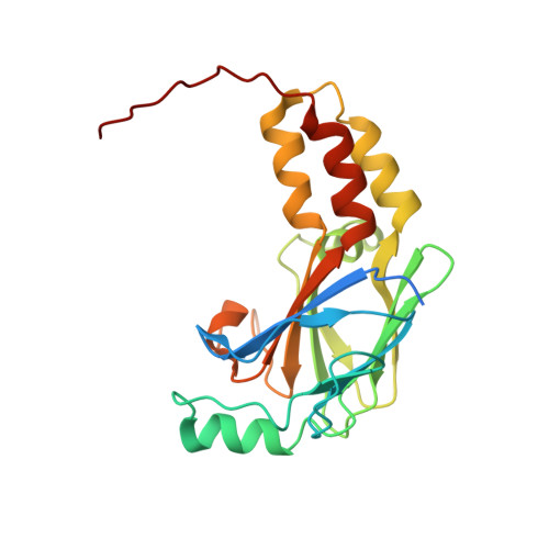
    1KHU

  • PubMed Abstract: 

    Phosphorylation of Smad1 at the conserved carboxyl terminal SVS sequence activates BMP signaling. Here we report the crystal structure of the Smad1 MH2 domain in a conformation that reveals the structural effects of phosphorylation and a molecular mechanism for activation. Within a trimeric subunit assembly, the SVS sequence docks near two putative phosphoserine binding pockets of the neighboring molecule, in a position ready to interact upon phosphorylation. The MH2 domain undergoes concerted conformational changes upon activation, which signal Smad1 dissociation from the receptor kinase for subsequent heteromeric assembly with Smad4. Biochemical and modeling studies reveal unique favorable interactions within the Smad1/Smad4 heteromeric interface, providing a structural basis for their association in signaling.


  • Organizational Affiliation
    • Department of Biochemistry and Molecular Pharmacology, University of Massachusetts Medical School, Worcester, MA 01655, USA.

Macromolecule Content 

  • Total Structure Weight: 98.18 kDa 
  • Atom Count: 6,772 
  • Modeled Residue Count: 776 
  • Deposited Residue Count: 872 
  • Unique protein chains: 1

Macromolecules

Find similar proteins by:|  3D Structure
Entity ID: 1
MoleculeChains  Sequence LengthOrganismDetailsImage
SMAD1
A, B, C, D
218Homo sapiensMutation(s): 0 
UniProt & NIH Common Fund Data Resources
Find proteins for Q15797 (Homo sapiens)
Explore Q15797 
Go to UniProtKB:  Q15797
PHAROS:  Q15797
GTEx:  ENSG00000170365 
Entity Groups
Sequence Clusters30% Identity50% Identity70% Identity90% Identity95% Identity100% Identity
UniProt GroupQ15797
Sequence Annotations
Expand
Reference Sequence

Experimental Data & Validation

Experimental Data

  • Method: X-RAY DIFFRACTION
  • Resolution: 2.50 Å
  • R-Value Free:  0.274 (Depositor), 0.271 (DCC) 
  • R-Value Work:  0.239 (Depositor), 0.240 (DCC) 
Space Group: H 3
Unit Cell:
Length ( Å )Angle ( ˚ )
a = 138.129α = 90
b = 138.129β = 90
c = 199.34γ = 120
Software Package:
Software NamePurpose
DENZOdata reduction
SCALAdata scaling
CNSrefinement
CCP4data scaling
CNSphasing

Structure Validation

View Full Validation Report


Entry History 

Deposition Data

Revision History  (Full details and data files)

  • Version 1.0: 2001-12-12
    Type: Initial release
  • Version 1.1: 2008-04-27
    Changes: Version format compliance
  • Version 1.2: 2011-07-13
    Changes: Version format compliance
  • Version 1.3: 2024-02-14
    Changes: Data collection, Database references
  • Version 1.4: 2024-04-03
    Changes: Refinement description