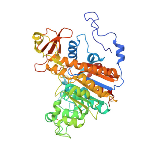
Artificial evolution of an enzyme active site: structural studies of three highly active mutants of Escherichia coli alkaline phosphatase.
Le Du, M.H., Lamoure, C., Muller, B.H., Bulgakov, O.V., Lajeunesse, E., Menez, A., Boulain, J.C.(2002) J Mol Biology 316: 941-953
- PubMed: 11884134
- DOI: https://doi.org/10.1006/jmbi.2001.5384
- Primary Citation of Related Structures:
1KH4, 1KH5, 1KH7, 1KH9, 1KHJ, 1KHK, 1KHL, 1KHN - PubMed Abstract:
The crystal structure of three mutants of Escherichia coli alkaline phosphatase with catalytic activity (k(cat)) enhancement as compare to the wild-type enzyme is described in different states. The biological aspects of this study have been reported elsewhere. The structure of the first mutant, D330N, which is threefold more active than the wild-type enzyme, was determined with phosphate in the active site, or with aluminium fluoride, which mimics the transition state. These structures reveal, in particular, that this first mutation does not alter the active site. The second mutant, D153H-D330N, is 17-fold more active than the wild-type enzyme and activated by magnesium, but its activity drops after few days. The structure of this mutant was solved under four different conditions. The phosphate-free enzyme was studied in an inactivated form with zinc at site M3, or after activation by magnesium. The comparison of these two forms free of phosphate illustrates the mechanism of the magnesium activation of the catalytic serine residue. In the presence of magnesium, the structure was determined with phosphate, or aluminium fluoride. The drop in activity of the mutant D153H-D330N could be explained by the instability of the metal ion at M3. The analysis of this mutant helped in the design of the third mutant, D153G-D330N. This mutant is up to 40-fold more active than the wild-type enzyme, with a restored robustness of the enzyme stability. The structure is presented here with covalently bound phosphate in the active site, representing the first phosphoseryl intermediate of a highly active alkaline phosphatase. This study shows how structural analysis may help to progress in the improvement of an enzyme catalytic activity (k(cat)), and explains the structural events associated with this artificial evolution.
- Département d'Ingénierie et d'Etudes des Protéines, CEA, Saclay, Gif-sur-Yvette, France. mhledu@cea.fr
Organizational Affiliation:


