1KFX | pdb_00001kfx

Crystal Structure of Human m-Calpain Form I


Experimental Data Snapshot

  • Method: X-RAY DIFFRACTION
  • Resolution: 3.15 Å
  • R-Value Free: 
    0.318 (Depositor) 
  • R-Value Work: 
    0.243 (Depositor) 

Starting Model: experimental
View more details

wwPDB Validation 3D Report Full Report

Validation slider image for 1KFX

This is version 1.4 of the entry. See complete history

Literature

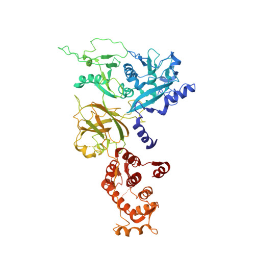
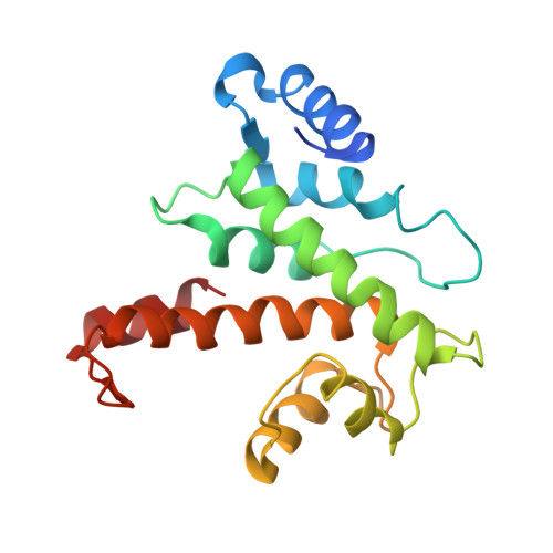
The crystal structure of calcium-free human m-calpain suggests an electrostatic switch mechanism for activation by calcium.

Strobl, S.Fernandez-Catalan, C.Braun, M.Huber, R.Masumoto, H.Nakagawa, K.Irie, A.Sorimachi, H.Bourenkow, G.Bartunik, H.Suzuki, K.Bode, W.

(2000) Proc Natl Acad Sci U S A 97: 588-592

  • DOI: https://doi.org/10.1073/pnas.97.2.588
  • Primary Citation Related Structures: 
    1KFU, 1KFX

  • PubMed Abstract: 

    Calpains (calcium-dependent cytoplasmic cysteine proteinases) are implicated in processes such as cytoskeleton remodeling and signal transduction. The 2.3-A crystal structure of full-length heterodimeric [80-kDa (dI-dIV) + 30-kDa (dV+dVI)] human m-calpain crystallized in the absence of calcium reveals an oval disc-like shape, with the papain-like catalytic domain dII and the two calmodulin-like domains dIV+dVI occupying opposite poles, and the tumor necrosis factor alpha-like beta-sandwich domain dIII and the N-terminal segments dI+dV located between. Compared with papain, the two subdomains dIIa+dIIb of the catalytic unit are rotated against one another by 50 degrees, disrupting the active site and the substrate binding site, explaining the inactivity of calpains in the absence of calcium. Calcium binding to an extremely negatively charged loop of domain dIII (an electrostatic switch) could release the adjacent barrel-like subdomain dIIb to move toward the helical subdomain dIIa, allowing formation of a functional catalytic center. This switch loop could also mediate membrane binding, thereby explaining calpains' strongly reduced calcium requirements in vivo. The activity status at the catalytic center might be further modulated by calcium binding to the calmodulin domains via the N-terminal linkers.


  • Organizational Affiliation
    • Max-Planck-Institute of Biochemistry, Am Klopferspitz 18a, D 82 152 Planegg-Martinsried, Germany.

Macromolecule Content 

  • Total Structure Weight: 101.23 kDa 
  • Atom Count: 6,825 
  • Modeled Residue Count: 816 
  • Deposited Residue Count: 883 
  • Unique protein chains: 2

Macromolecules

Find similar proteins by:|  3D Structure
Entity ID: 1
MoleculeChains  Sequence LengthOrganismDetailsImage
M-CALPAIN LARGE SUBUNITA [auth L]699Homo sapiensMutation(s): 0 
EC: 3.4.22.53
UniProt & NIH Common Fund Data Resources
Find proteins for P17655 (Homo sapiens)
Explore P17655 
Go to UniProtKB:  P17655
PHAROS:  P17655
GTEx:  ENSG00000162909 
Entity Groups
Sequence Clusters30% Identity50% Identity70% Identity90% Identity95% Identity100% Identity
UniProt GroupP17655
Sequence Annotations
Expand
Reference Sequence
Find similar proteins by:|  3D Structure
Entity ID: 2
MoleculeChains  Sequence LengthOrganismDetailsImage
M-CALPAIN SMALL SUBUNITB [auth S]184Homo sapiensMutation(s): 0 
UniProt & NIH Common Fund Data Resources
Find proteins for P04632 (Homo sapiens)
Explore P04632 
Go to UniProtKB:  P04632
PHAROS:  P04632
GTEx:  ENSG00000126247 
Entity Groups
Sequence Clusters30% Identity50% Identity70% Identity90% Identity95% Identity100% Identity
UniProt GroupP04632
Sequence Annotations
Expand
Reference Sequence

Experimental Data & Validation

Experimental Data

  • Method: X-RAY DIFFRACTION
  • Resolution: 3.15 Å
  • R-Value Free:  0.318 (Depositor) 
  • R-Value Work:  0.243 (Depositor) 
Space Group: P 1 21 1
Unit Cell:
Length ( Å )Angle ( ˚ )
a = 64.78α = 90
b = 133.25β = 102.07
c = 77.53γ = 90
Software Package:
Software NamePurpose
MAINmodel building
CNSrefinement
DENZOdata reduction
CCP4data scaling
MAINphasing

Structure Validation

View Full Validation Report


Entry History 

Revision History  (Full details and data files)

  • Version 1.0: 2001-12-07
    Type: Initial release
  • Version 1.1: 2008-04-27
    Changes: Version format compliance
  • Version 1.2: 2011-07-13
    Changes: Version format compliance
  • Version 1.3: 2012-05-02
    Changes: Structure summary
  • Version 1.4: 2023-08-16
    Changes: Data collection, Database references, Refinement description