1KFR | pdb_00001kfr

Structural plasticity in the eight-helix fold of a trematode hemoglobin


Experimental Data Snapshot

  • Method: X-RAY DIFFRACTION
  • Resolution: 1.85 Å
  • R-Value Free: 
    0.220 (Depositor), 0.212 (DCC) 
  • R-Value Work: 
    0.161 (Depositor) 
  • R-Value Observed: 
    0.164 (Depositor) 

wwPDB Validation 3D Report Full Report

Validation slider image for 1KFR

Ligand Structure Quality Assessment 


This is version 1.3 of the entry. See complete history

Literature

Structural plasticity in the eight-helix fold of a trematode haemoglobin.

Milani, M.Pesce, A.Dewilde, S.Ascenzi, P.Moens, L.Bolognesi, M.

(2002) Acta Crystallogr D Biol Crystallogr 58: 719-722

  • DOI: https://doi.org/10.1107/s0907444902001865
  • Primary Citation Related Structures: 
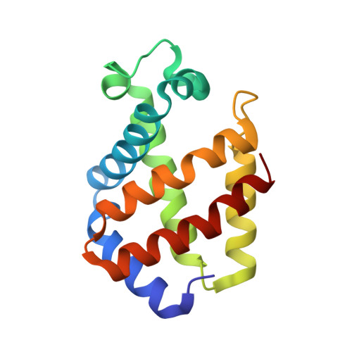
    1KFR

  • PubMed Abstract: 

    The three-dimensional structure of recombinant haemoglobin from the trematode Paramphistomum epiclitum, displaying the highest oxygen affinity so far observed for (non)vertebrate haemoglobins, has previously been determined at 1.17 A resolution (orthorhombic space group P2(1)2(1)2(1)). In the present communication, the three-dimensional structure of wild-type P. epiclitum haemoglobin is reported at 1.85 A resolution in a monoclinic crystal form (R factor = 16.1%, R(free) = 22.0%). Comparison of P. epiclitum (recombinant versus wild-type ferric Hb) structures in the two crystal forms shows structural differences in the haem proximal and distal sites which have not been reported for other known haemoglobin structures previously.


  • Organizational Affiliation
    • Department of Physics-INFM, Advanced Biotechnology Centre, University of Genova, Largo Rosanna Benzi 10, I-16146 Genova, Italy.

Macromolecule Content 

  • Total Structure Weight: 17.47 kDa 
  • Atom Count: 1,404 
  • Modeled Residue Count: 147 
  • Deposited Residue Count: 147 
  • Unique protein chains: 1

Macromolecules

Find similar proteins by:|  3D Structure
Entity ID: 1
MoleculeChains  Sequence LengthOrganismDetailsImage
Hemoglobin147Paramphistomum epiclitumMutation(s): 0 
UniProt
Find proteins for P80721 (Paramphistomum epiclitum)
Explore P80721 
Go to UniProtKB:  P80721
Entity Groups
Sequence Clusters30% Identity50% Identity70% Identity90% Identity95% Identity100% Identity
UniProt GroupP80721
Sequence Annotations
Expand
Reference Sequence

Experimental Data & Validation

Experimental Data

  • Method: X-RAY DIFFRACTION
  • Resolution: 1.85 Å
  • R-Value Free:  0.220 (Depositor), 0.212 (DCC) 
  • R-Value Work:  0.161 (Depositor) 
  • R-Value Observed: 0.164 (Depositor) 
Space Group: P 1 21 1
Unit Cell:
Length ( Å )Angle ( ˚ )
a = 41.117α = 90
b = 31.45β = 95.5
c = 54.952γ = 90
Software Package:
Software NamePurpose
EPMRphasing
REFMACrefinement
DENZOdata reduction
SCALEPACKdata scaling

Structure Validation

View Full Validation Report


Ligand Structure Quality Assessment 


Entry History 

Deposition Data

Revision History  (Full details and data files)

  • Version 1.0: 2002-04-24
    Type: Initial release
  • Version 1.1: 2008-04-27
    Changes: Version format compliance
  • Version 1.2: 2011-07-13
    Changes: Version format compliance
  • Version 1.3: 2024-02-07
    Changes: Data collection, Database references, Derived calculations