1KC4

NMR Structural Analysis of the Complex Formed Between alpha-Bungarotoxin and the Principal alpha-Neurotoxin Binding Sequence on the alpha7 Subunit of a Neuronal Nicotinic Acetylcholine Receptor


Experimental Data Snapshot

  • Method: SOLUTION NMR
  • Conformers Submitted: 10 
  • Selection Criteria: The submitted conformer models are the 10 with the lowest energy from a pool of over 100 accepted structures that had no restraint violations. 

wwPDB Validation   3D Report Full Report


This is version 1.3 of the entry. See complete history


Literature

NMR structural analysis of alpha-bungarotoxin and its complex with the principal alpha-neurotoxin-binding sequence on the alpha 7 subunit of a neuronal nicotinic acetylcholine receptor.

Moise, L.Piserchio, A.Basus, V.J.Hawrot, E.

(2002) J Biol Chem 277: 12406-12417

  • DOI: https://doi.org/10.1074/jbc.M110320200
  • Primary Citation of Related Structures:  
    1KC4, 1KFH, 1KL8

  • PubMed Abstract: 

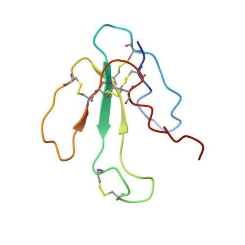
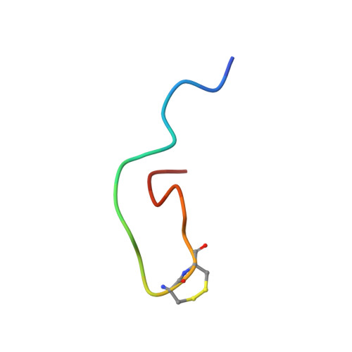
    We report a new, higher resolution NMR structure of alpha-bungarotoxin that defines the structure-determining disulfide core and beta-sheet regions. We further report the NMR structure of the stoichiometric complex formed between alpha-bungarotoxin and a recombinantly expressed 19-mer peptide ((178)IPGKRTESFYECCKEPYPD(196)) derived from the alpha7 subunit of the chick neuronal nicotinic acetylcholine receptor. A comparison of these two structures reveals binding-induced stabilization of the flexible tip of finger II in alpha-bungarotoxin. The conformational rearrangements in the toxin create an extensive binding surface involving both sides of the alpha7 19-mer hairpin-like structure. At the contact zone, Ala(7), Ser(9), and Ile(11) in finger I and Arg(36), Lys(38), Val(39), and Val(40) in finger II of alpha-bungarotoxin interface with Phe(186), Tyr(187), Glu(188), and Tyr(194) in the alpha7 19-mer underscoring the importance of receptor aromatic residues as critical neurotoxin-binding determinants. Superimposing the structure of the complex onto that of the acetylcholine-binding protein (1I9B), a soluble homologue of the extracellular domain of the alpha7 receptor, places alpha-bungarotoxin at the peripheral surface of the inter-subunit interface occluding the agonist-binding site. The disulfide-rich core of alpha-bungarotoxin is suggested to be tilted in the direction of the membrane surface with finger II extending into the proposed ligand-binding cavity.


  • Organizational Affiliation

    Department of Molecular Pharmacology, Brown University, Providence, Rhode Island 02912, USA.


Macromolecules
Find similar proteins by:  (by identity cutoff)  |  3D Structure
Entity ID: 1
MoleculeChains Sequence LengthOrganismDetailsImage
alpha-bungarotoxin74Bungarus multicinctusMutation(s): 0 
UniProt
Find proteins for P60615 (Bungarus multicinctus)
Explore P60615 
Go to UniProtKB:  P60615
Entity Groups  
Sequence Clusters30% Identity50% Identity70% Identity90% Identity95% Identity100% Identity
UniProt GroupP60615
Sequence Annotations
Expand
  • Reference Sequence

Find similar proteins by:  Sequence   |   3D Structure  

Entity ID: 2
MoleculeChains Sequence LengthOrganismDetailsImage
neuronal acetylcholine receptor protein, alpha-7 chain20Gallus gallusMutation(s): 1 
UniProt
Find proteins for P22770 (Gallus gallus)
Explore P22770 
Go to UniProtKB:  P22770
Entity Groups  
Sequence Clusters30% Identity50% Identity70% Identity90% Identity95% Identity100% Identity
UniProt GroupP22770
Sequence Annotations
Expand
  • Reference Sequence
Small Molecules
Modified Residues  1 Unique
IDChains TypeFormula2D DiagramParent
HSL
Query on HSL
B
L-PEPTIDE LINKINGC4 H7 N O2SER
Experimental Data & Validation

Experimental Data

  • Method: SOLUTION NMR
  • Conformers Submitted: 10 
  • Selection Criteria: The submitted conformer models are the 10 with the lowest energy from a pool of over 100 accepted structures that had no restraint violations. 

Structure Validation

View Full Validation Report


Entry History 

Deposition Data

Revision History  (Full details and data files)

  • Version 1.0: 2002-03-13
    Type: Initial release
  • Version 1.1: 2008-04-27
    Changes: Version format compliance
  • Version 1.2: 2011-07-13
    Changes: Version format compliance
  • Version 1.3: 2021-10-27
    Changes: Data collection, Database references, Derived calculations