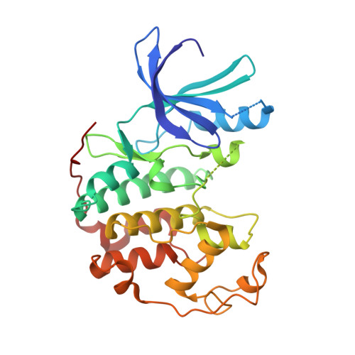
1JVP

Crystal structure of human CDK2 (unphosphorylated) in complex with PKF049-365


Experimental Data Snapshot

  • Method: X-RAY DIFFRACTION
  • Resolution: 1.53 Å
  • R-Value Free: 0.253 
  • R-Value Work: 0.206 
  • R-Value Observed: 0.210 

wwPDB Validation   3D Report Full Report


Ligand Structure Quality Assessment 


This is version 2.1 of the entry. See complete history


Literature

Structure-based design and protein X-ray analysis of a protein kinase inhibitor.

Furet, P.Meyer, T.Strauss, A.Raccuglia, S.Rondeau, J.M.

(2002) Bioorg Med Chem Lett 12: 221-224

  • DOI: https://doi.org/10.1016/s0960-894x(01)00715-6
  • Primary Citation of Related Structures:  
    1JVP

  • PubMed Abstract: 

    A 5-aryl-1H-pyrazole molecular scaffold was designed to ligate the ATP binding site of cyclin dependent kinase 2 (CDK2) on the basis of crystallographic information. A search of the compound collection of Novartis using this scaffold as substructure query led to the identification of PKF049-365 as a representative of a new class of inhibitors of the cell cycle kinases CDK1/2. The three-dimensional structure of CDK2 in complex with PKF049-365 was subsequently determined by protein crystallography and refined to 1.53 A resolution. The X-ray analysis confirmed the binding mode expected from the design hypothesis. In addition, it revealed an alternative binding orientation involving a second tautomeric form of the inhibitor that was not envisaged during the design stage.


  • Organizational Affiliation

    Novartis Pharmaceuticals Inc., Oncology Research, CH-4002 Basel, Switzerland. pascal.furet@pharma.novartis.com


Macromolecules
Find similar proteins by:  (by identity cutoff)  |  3D Structure
Entity ID: 1
MoleculeChains Sequence LengthOrganismDetailsImage
Cell division protein kinase 2A [auth P]298Homo sapiensMutation(s): 0 
EC: 2.7.1.37 (PDB Primary Data), 2.7.11.22 (UniProt)
UniProt & NIH Common Fund Data Resources
Find proteins for P24941 (Homo sapiens)
Explore P24941 
Go to UniProtKB:  P24941
PHAROS:  P24941
GTEx:  ENSG00000123374 
Entity Groups  
Sequence Clusters30% Identity50% Identity70% Identity90% Identity95% Identity100% Identity
UniProt GroupP24941
Sequence Annotations
Expand
  • Reference Sequence
Small Molecules
Ligands 1 Unique
IDChains Name / Formula / InChI Key2D Diagram3D Interactions
89E
Query on 89E

Download Ideal Coordinates CCD File 
B [auth P]3-pyridin-4-yl-2,4-dihydroindeno[1,2-c]pyrazole
C15 H11 N3
NHOACLCXCKJMAK-UHFFFAOYSA-N
Experimental Data & Validation

Experimental Data

  • Method: X-RAY DIFFRACTION
  • Resolution: 1.53 Å
  • R-Value Free: 0.253 
  • R-Value Work: 0.206 
  • R-Value Observed: 0.210 
  • Space Group: P 21 21 21
Unit Cell:
Length ( Å )Angle ( ˚ )
a = 53.528α = 90
b = 71.802β = 90
c = 72.362γ = 90
Software Package:
Software NamePurpose
AMoREphasing
X-PLORrefinement
DENZOdata reduction
SCALEPACKdata scaling

Structure Validation

View Full Validation Report


Ligand Structure Quality Assessment 


Entry History 

Deposition Data

Revision History  (Full details and data files)

  • Version 1.0: 2001-12-21
    Type: Initial release
  • Version 1.1: 2008-04-27
    Changes: Version format compliance
  • Version 1.2: 2011-07-13
    Changes: Version format compliance
  • Version 2.0: 2021-09-22
    Changes: Atomic model, Data collection, Database references, Derived calculations, Non-polymer description, Structure summary
  • Version 2.1: 2024-02-07
    Changes: Data collection