1JUJ | pdb_00001juj

Human Thymidylate Synthase Bound to dUMP and LY231514, a Pyrrolo(2,3-d)pyrimidine-based Antifolate


Experimental Data Snapshot

  • Method: X-RAY DIFFRACTION
  • Resolution: 3.00 Å
  • R-Value Free: 
    0.311 (Depositor), 0.308 (DCC) 
  • R-Value Work: 
    0.268 (Depositor), 0.268 (DCC) 
  • R-Value Observed: 
    0.273 (Depositor) 

Starting Model: experimental
View more details

wwPDB Validation 3D Report Full Report

Validation slider image for 1JUJ

Ligand Structure Quality Assessment 


This is version 1.5 of the entry. See complete history

Literature

Multi-targeted antifolates aimed at avoiding drug resistance form covalent closed inhibitory complexes with human and Escherichia coli thymidylate synthases.

Sayre, P.H.Finer-Moore, J.S.Fritz, T.A.Biermann, D.Gates, S.B.MacKellar, W.C.Patel, V.F.Stroud, R.M.

(2001) J Mol Biology 313: 813-829

  • DOI: https://doi.org/10.1006/jmbi.2001.5074
  • Primary Citation Related Structures: 
    1JTQ, 1JTU, 1JU6, 1JUJ, 1JUT

  • PubMed Abstract: 

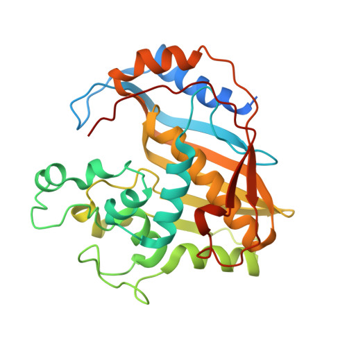
    Crystal structures of four pyrrolo(2,3-d)pyrimidine-based antifolate compounds, developed as inhibitors of thymidylate synthase (TS) in a strategy to circumvent drug-resistance, have been determined in complexes with their in vivo target, human thymidylate synthase, and with the structurally best-characterized Escherichia coli enzyme, to resolutions of 2.2-3.0 A. The 2.9 A crystal structure of a complex of human TS with one of the inhibitors, the multi-targeted antifolate LY231514, demonstrates that this compound induces a "closed" enzyme conformation and leads to formation of a covalent bond between enzyme and substrate. This structure is one of the first liganded human TS structures, and its solution was aided by mutation to facilitate crystallization. Structures of three other pyrrolo(2,3-d)pyrimidine-based antifolates in complex with Escherichia coli TS confirm the orientation of this class of inhibitors in the active site. Specific interactions between the polyglutamyl moiety and a positively charged groove on the enzyme surface explain the marked increase in affinity of the pyrrolo(2,3-d)pyrimidine inhibitors once they are polyglutamylated, as mediated in vivo by the cellular enzyme folyl polyglutamate synthetase.


  • Organizational Affiliation
    • Department of Biochemistry and Biophysics, University of California, San Francisco, CA 94143-0448, USA.

Macromolecule Content 

  • Total Structure Weight: 145.88 kDa 
  • Atom Count: 9,412 
  • Modeled Residue Count: 1,144 
  • Deposited Residue Count: 1,252 
  • Unique protein chains: 1

Macromolecules

Find similar proteins by:|  3D Structure
Entity ID: 1
MoleculeChains  Sequence LengthOrganismDetailsImage
THYMIDYLATE SYNTHASE
A, B, C, D
313Homo sapiensMutation(s): 1 
EC: 2.1.1.45
UniProt & NIH Common Fund Data Resources
Find proteins for P04818 (Homo sapiens)
Explore P04818 
Go to UniProtKB:  P04818
PHAROS:  P04818
GTEx:  ENSG00000176890 
Entity Groups
Sequence Clusters30% Identity50% Identity70% Identity90% Identity95% Identity100% Identity
UniProt GroupP04818
Sequence Annotations
Expand
Reference Sequence

Small Molecules

Ligands 2 Unique
IDChains Name / Formula / InChI Key2D Diagram3D Interactions
LYA

Query on LYA


Download:Ideal Coordinates CCD File
F [auth A],
H [auth B],
J [auth C],
L [auth D]
2-{4-[2-(2-AMINO-4-OXO-4,7-DIHYDRO-3H-PYRROLO[2,3-D]PYRIMIDIN-5-YL)-ETHYL]-BENZOYLAMINO}-PENTANEDIOIC ACID
C20 H21 N5 O6
WBXPDJSOTKVWSJ-ZDUSSCGKSA-N
UMP

Query on UMP


Download:Ideal Coordinates CCD File
E [auth A],
G [auth B],
I [auth C],
K [auth D]
2'-DEOXYURIDINE 5'-MONOPHOSPHATE
C9 H13 N2 O8 P
JSRLJPSBLDHEIO-SHYZEUOFSA-N

Experimental Data & Validation

Experimental Data

  • Method: X-RAY DIFFRACTION
  • Resolution: 3.00 Å
  • R-Value Free:  0.311 (Depositor), 0.308 (DCC) 
  • R-Value Work:  0.268 (Depositor), 0.268 (DCC) 
  • R-Value Observed: 0.273 (Depositor) 
Space Group: C 1 2 1
Unit Cell:
Length ( Å )Angle ( ˚ )
a = 138.14α = 90
b = 98.14β = 110.7
c = 111.97γ = 90
Software Package:
Software NamePurpose
DENZOdata reduction
SCALEPACKdata scaling
CNSrefinement
CNSphasing

Structure Validation

View Full Validation Report


Ligand Structure Quality Assessment 


Entry History 

Deposition Data

Revision History  (Full details and data files)

  • Version 1.0: 2001-09-19
    Type: Initial release
  • Version 1.1: 2008-04-27
    Changes: Version format compliance
  • Version 1.2: 2011-07-13
    Changes: Version format compliance
  • Version 1.3: 2021-10-27
    Changes: Database references, Derived calculations
  • Version 1.4: 2024-04-03
    Changes: Data collection, Refinement description
  • Version 1.5: 2024-10-30
    Changes: Structure summary