1JTP

Degenerate interfaces in antigen-antibody complexes


Experimental Data Snapshot

  • Method: X-RAY DIFFRACTION
  • Resolution: 1.90 Å
  • R-Value Free: 0.229 
  • R-Value Work: 0.184 

wwPDB Validation   3D Report Full Report


This is version 1.4 of the entry. See complete history


Literature

Degenerate interfaces in antigen-antibody complexes.

Decanniere, K.Transue, T.R.Desmyter, A.Maes, D.Muyldermans, S.Wyns, L.

(2001) J Mol Biol 313: 473-478

  • DOI: https://doi.org/10.1006/jmbi.2001.5075
  • Primary Citation of Related Structures:  
    1JTO, 1JTP, 1JTT

  • PubMed Abstract: 

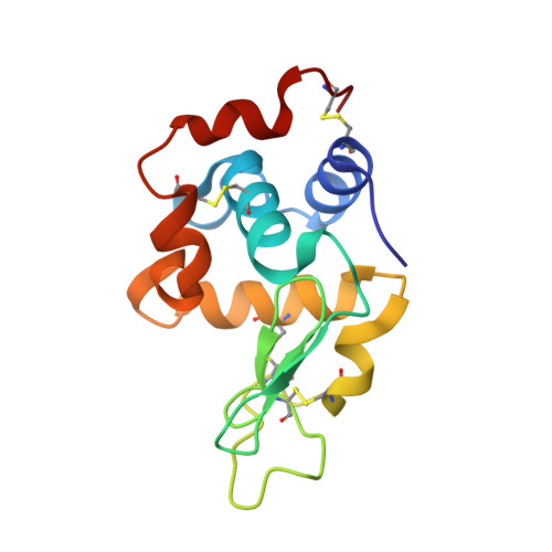
    In most of the work dealing with the analysis of protein-protein interfaces, a single X-ray structure is available or selected, and implicitly it is assumed that this structure corresponds to the optimal complex for this pair of proteins. However, we have found a degenerate interface in a high-affinity antibody-antigen complex: the two independent complexes of the camel variable domain antibody fragment cAb-Lys3 and its antigen hen egg white lysozyme present in the asymmetric unit of our crystals show a difference in relative orientation between antibody and antigen, leading to important differences at the protein-protein interface. A third cAb-Lys3-hen lysozyme complex in a different crystal form adopts yet another relative orientation. Our results show that protein-protein interface characteristics can vary significantly between different specimens of the same high-affinity antibody-protein antigen complex. Consideration should be given to this type of observation when trying to establish general protein-protein interface characteristics.


  • Organizational Affiliation

    Vrije Universiteit Brussel Dienst Ultrastructuur, Vlaams Instituut voor Biotechnologie, Paardenstraat 65, B-1640 St.-Genesius Rode, Belgium. klaas@ultr.vub.ac.be


Macromolecules
Find similar proteins by:  (by identity cutoff)  |  3D Structure
Entity ID: 1
MoleculeChains Sequence LengthOrganismDetailsImage
Single-Domain Antibody
A, B
148Camelus dromedariusMutation(s): 0 
Entity Groups  
Sequence Clusters30% Identity50% Identity70% Identity90% Identity95% Identity100% Identity
Sequence Annotations
Expand
  • Reference Sequence
Find similar proteins by:  (by identity cutoff)  |  3D Structure
Entity ID: 2
MoleculeChains Sequence LengthOrganismDetailsImage
LYSOZYME CC [auth L],
D [auth M]
129Meleagris gallopavoMutation(s): 0 
EC: 3.2.1.17
UniProt
Find proteins for P00703 (Meleagris gallopavo)
Explore P00703 
Go to UniProtKB:  P00703
Entity Groups  
Sequence Clusters30% Identity50% Identity70% Identity90% Identity95% Identity100% Identity
UniProt GroupP00703
Sequence Annotations
Expand
  • Reference Sequence
Small Molecules
Ligands 2 Unique
IDChains Name / Formula / InChI Key2D Diagram3D Interactions
FMT
Query on FMT

Download Ideal Coordinates CCD File 
F [auth A]
H [auth B]
K [auth L]
M
N [auth M]
F [auth A],
H [auth B],
K [auth L],
M,
N [auth M],
O [auth M]
FORMIC ACID
C H2 O2
BDAGIHXWWSANSR-UHFFFAOYSA-N
NA
Query on NA

Download Ideal Coordinates CCD File 
E [auth A],
G [auth B],
I [auth L],
J [auth L],
L [auth M]
SODIUM ION
Na
FKNQFGJONOIPTF-UHFFFAOYSA-N
Experimental Data & Validation

Experimental Data

  • Method: X-RAY DIFFRACTION
  • Resolution: 1.90 Å
  • R-Value Free: 0.229 
  • R-Value Work: 0.184 
  • Space Group: P 21 21 21
Unit Cell:
Length ( Å )Angle ( ˚ )
a = 67.123α = 90
b = 69.375β = 90
c = 113.231γ = 90
Software Package:
Software NamePurpose
MAR345data collection
SCALEPACKdata scaling
CNSrefinement
CNSphasing

Structure Validation

View Full Validation Report


Entry History 

Deposition Data

Revision History  (Full details and data files)

  • Version 1.0: 2001-12-05
    Type: Initial release
  • Version 1.1: 2008-04-27
    Changes: Version format compliance
  • Version 1.2: 2011-07-13
    Changes: Version format compliance
  • Version 1.3: 2017-10-04
    Changes: Refinement description
  • Version 1.4: 2023-08-16
    Changes: Data collection, Database references, Derived calculations, Refinement description