1JS4

ENDO/EXOCELLULASE:CELLOBIOSE FROM THERMOMONOSPORA


Experimental Data Snapshot

  • Method: X-RAY DIFFRACTION
  • Resolution: 2.00 Å
  • R-Value Work: 0.199 
  • R-Value Observed: 0.199 

wwPDB Validation   3D Report Full Report


Ligand Structure Quality Assessment 


This is version 2.0 of the entry. See complete history


Literature

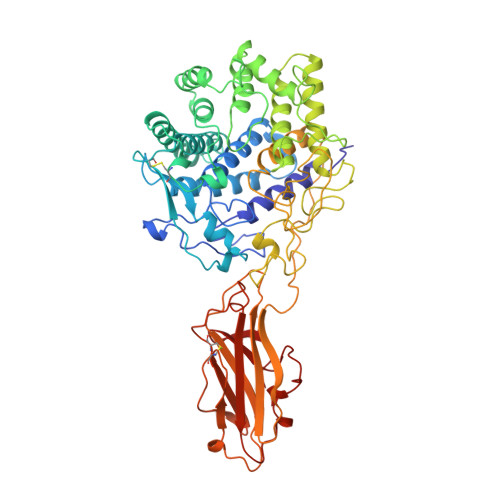
Structure and mechanism of endo/exocellulase E4 from Thermomonospora fusca.

Sakon, J.Irwin, D.Wilson, D.B.Karplus, P.A.

(1997) Nat Struct Biol 4: 810-818

  • DOI: https://doi.org/10.1038/nsb1097-810
  • Primary Citation of Related Structures:  
    1JS4, 1TF4, 3TF4, 4TF4

  • PubMed Abstract: 

    Cellulase E4 from Thermomonospora fusca is unusual in that it has characteristics of both exo- and endo-cellulases. Here we report the crystal structure of a 68K M(r) fragment of E4 (E4-68) at 1.9 A resolution. E4-68 contains both a family 9 catalytic domain, exhibiting an (alpha/alpha)6 barrel fold, and a family III cellulose binding domain, having an antiparallel beta-sandwich fold. While neither of these folds is novel, E4-68 provides the first cellulase structure having interacting catalytic and cellulose binding domains. The complexes of E4-68 with cellopentaose, cellotriose and cellobiose reveal conformational changes associated with ligand binding and allow us to propose a catalytic mechanism for family 9 enzymes. We also provide evidence that E4 has two novel characteristics: first it combines exo- and endo-activities and second, when it functions as an exo-cellulase, it cleaves off cellotetraose units.


  • Organizational Affiliation

    Section of Biochemistry, Molecular and Cell Biology, Cornell University, Ithaca, New York 14853, USA.


Macromolecules
Find similar proteins by:  (by identity cutoff)  |  3D Structure
Entity ID: 1
MoleculeChains Sequence LengthOrganismDetailsImage
ENDO/EXOCELLULASE E4
A, B
605Thermobifida fuscaMutation(s): 0 
Gene Names: BAMH1-PST1 FRAGMENT OF T. FUSC
EC: 3.2.1.4
UniProt
Find proteins for P26221 (Thermobifida fusca)
Explore P26221 
Go to UniProtKB:  P26221
Entity Groups  
Sequence Clusters30% Identity50% Identity70% Identity90% Identity95% Identity100% Identity
UniProt GroupP26221
Sequence Annotations
Expand
  • Reference Sequence
Oligosaccharides

Help

Entity ID: 2
MoleculeChains Length2D Diagram Glycosylation3D Interactions
beta-D-glucopyranose-(1-4)-beta-D-glucopyranose-(1-4)-beta-D-glucopyranose
C
3N/A
Glycosylation Resources
GlyTouCan:  G14338VK
GlyCosmos:  G14338VK
GlyGen:  G14338VK
Entity ID: 3
MoleculeChains Length2D Diagram Glycosylation3D Interactions
beta-D-glucopyranose-(1-4)-beta-D-glucopyranose
D
2N/A
Glycosylation Resources
GlyTouCan:  G84824ZO
GlyCosmos:  G84824ZO
Biologically Interesting Molecules (External Reference) 2 Unique
Experimental Data & Validation

Experimental Data

  • Method: X-RAY DIFFRACTION
  • Resolution: 2.00 Å
  • R-Value Work: 0.199 
  • R-Value Observed: 0.199 
  • Space Group: P 41 21 2
Unit Cell:
Length ( Å )Angle ( ˚ )
a = 145.68α = 90
b = 145.68β = 90
c = 157.08γ = 90
Software Package:
Software NamePurpose
SCALEPACKdata scaling
PHASESphasing
X-PLORmodel building
X-PLORrefinement
X-PLORphasing

Structure Validation

View Full Validation Report


Ligand Structure Quality Assessment 


Entry History 

Deposition Data

Revision History  (Full details and data files)

  • Version 1.0: 1997-09-17
    Type: Initial release
  • Version 1.1: 2008-03-24
    Changes: Version format compliance
  • Version 1.2: 2011-07-13
    Changes: Version format compliance
  • Version 2.0: 2020-07-29
    Type: Remediation
    Reason: Carbohydrate remediation
    Changes: Advisory, Atomic model, Data collection, Derived calculations, Structure summary