Crystal structures of Escherichia coli dihydrofolate reductase complexed with 5-formyltetrahydrofolate (folinic acid) in two space groups: evidence for enolization of pteridine O4.
Lee, H., Reyes, V.M., Kraut, J.(1996) Biochemistry 35: 7012-7020
- PubMed: 8679526 Search on PubMed
- DOI: https://doi.org/10.1021/bi960028g
- Primary Citation Related Structures:
1JOL, 1JOM - PubMed Abstract:
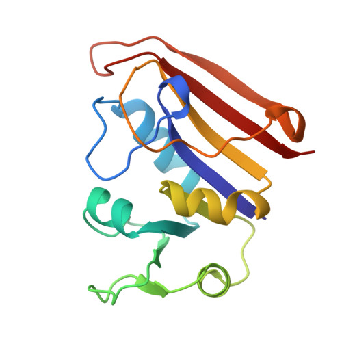
The crystal structure of Escherichia coli dihydrofolate reductase (ecDHFR, EC 1.5.1.3) as a binary complex with folinic acid (5-formyl-5,6,7,8-tetrahydrofolate; also called leucovorin or citrovorum factor) has been solved in two space groups, P6(1) and P6(5), with, respectively, two molecules and one molecule per asymmetric unit. The crystal structures have been refined to an R-factor of 14.2% at resolutions of 2.0 and 1.9 A. The P6(1) structure is isomorphous with several previously reported ecDHFR binary complexes [Bolin, J.T., Filman, D.J., Matthews, D.A., Hamlin, R.C., & Kraut, J. (1982) J. Biol. Chem. 257, 13650-13662; Reyes, V.M., Sawaya, M.R., Brown, K.A., & Kraut, J. (1995) Biochemistry 34, 2710-2723]; enzyme and ligand conformations are very similar to the P6(1) 5,10-dideazatetrahydrofolate complex. While the two enzyme subdomains of the P6(1) structure are nearly in the closed conformation, exemplified by the methotrexate P6(1) binary complex, in the P6(5) structure they are in an intermediate conformation, halfway between the closed and the fully open conformation of the apoenzyme [Bystroff, C., Oatley, S.J., & Kraut, J. (1990) Biochemistry 29, 3263-3277]. Thus crystal packing strongly influences this aspect of the enzyme structure. In contrast to the P6(1) structure, in which the Met-20 loop (residues 9-23) is turned away from the substrate binding pocket, in the P6(5) structure the Met-20 loop blocks the pocket and protrudes into the cofactor binding site. In this respect, the P6(5) structure is unique. Additionally, positioning of a Ca2+ ion (a component of the crystallization medium) is different in the two crystal packings: in the P6(1) structure it lies at the boundary between the two molecules of the asymmetric unit, while in P6(5) it coordinates two water molecules, the hydroxyl group of an ethanol molecule, and the backbone carbonyl oxygens of Glu-17, Asn-18, and Met-20. The Ca2+ ion thus stabilizes a single turn of 3(10) helix (residues 16-18 in the Met-20 loop), a second unique feature of the P6(5) crystal structure. The disposition of the N5-formyl group in these structures indicates formation, at least half of the time, of an intramolecular hydrogen bond between the formyl oxygen and O4 of the tetrahydropterin ring. This observation is consistent with the existence of an enol-keto equilibrium in which the enolic tautomer is favored when a hydrogen-bond acceptor is present between O4 and N5. Such would be the case whenever a water molecule occupies that site as part of a hypothetical proton-relay mechanism. Two arginine side chains, Arg-52 in the P6(5) structure and Arg-44 in molecule A of the P6(1) structure, are turned away drastically from the ligand (p-aminobenzoyl)glutamic acid moiety as compared with previously reported DHFR binary complex structures. As in the ecDHFR dideazatetrahydrofolate complex, in both the P6(1) and P6(5) structures a water molecule bridges pteridine O4 and Trp-22(N epsilon 1) with ideal geometry for hydrogen bonding, perhaps contributing to the slow release of 5,6,7,8-tetrahydrofolate from the enzyme-product complex. When either the P6(1) or the P6(5) structures are superimposed with the NADPH holoenzyme [Sawaya, M. R. (1994) Ph.D. Dissertation, University of California, San Diego], we find that the distances between the nicotinamide C4 and pteridine C6 and C7 are very short, 2.1 and 1.7 A in the P6(1) case and 2.0 and 1.4 A in the P6(5) case, perhaps in part explaining the more rapid release of tetrahydrofolate from the enzyme-product complex when NADPH is bound.
- Department of Chemistry and Biochemistry, University of California, San Diego, La Jolla 92093-0506, USA.
Organizational Affiliation: