1JJ1

CRYSTAL STRUCTURE OF ORTHORHOMBIC LYSOZYME GROWN AT PH 4.6 IN PRESENCE OF 5% SORBITOL


Experimental Data Snapshot

  • Method: X-RAY DIFFRACTION
  • Resolution: 1.90 Å
  • R-Value Free: 0.223 
  • R-Value Work: 0.186 
  • R-Value Observed: 0.186 

wwPDB Validation   3D Report Full Report


This is version 1.4 of the entry. See complete history


Literature

The effect of stabilizing additives on the structure and hydration of proteins: a study involving tetragonal lysozyme.

Datta, S.Biswal, B.K.Vijayan, M.

(2001) Acta Crystallogr D Biol Crystallogr 57: 1614-1620

  • DOI: https://doi.org/10.1107/s090744490101280x
  • Primary Citation of Related Structures:  
    1JIS, 1JIT, 1JIY, 1JJ0, 1JJ1, 1JJ3

  • PubMed Abstract: 

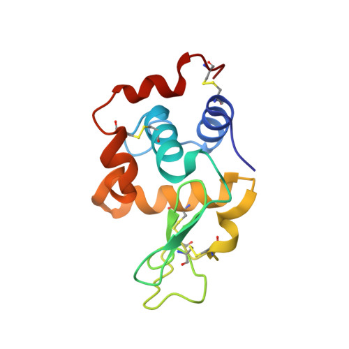
    In order to elucidate the effect of stabilizing additives on the structure of proteins and the associated ordered water molecules in the hydration shell, the crystal structures of tetragonal lysozyme grown in the presence of sucrose, sorbitol and trehalose have been refined. Also refined are the structures of orthorhombic and monoclinic lysozyme grown under the conditions in which tetragonal lysozyme is normally grown. A comparison of the two sets of structures with the structure of native tetragonal lysozyme shows that the effect of the additives on the structure of the protein molecule is less than that of the normal minor changes associated with differences in molecular packing. Surprisingly, the same is true of the effect on the hydration shell, represented by the ordered water molecules attached to the protein. Thus, it appears that the cause of the stabilizing effect of the additives needs to be sought outside the immediate neighbourhood of the protein molecule. Sorbitol and trehalose do not coherently interact with the protein. One sucrose molecule binds at the active-site cleft of the enzyme.


  • Organizational Affiliation

    Molecular Biophysics Unit, Indian Institute of Science, Bangalore 560 012, India.


Macromolecules
Find similar proteins by:  (by identity cutoff)  |  3D Structure
Entity ID: 1
MoleculeChains Sequence LengthOrganismDetailsImage
LYSOZYME129Gallus gallusMutation(s): 0 
EC: 3.2.1.17
UniProt
Find proteins for P00698 (Gallus gallus)
Explore P00698 
Go to UniProtKB:  P00698
Entity Groups  
Sequence Clusters30% Identity50% Identity70% Identity90% Identity95% Identity100% Identity
UniProt GroupP00698
Sequence Annotations
Expand
  • Reference Sequence
Experimental Data & Validation

Experimental Data

  • Method: X-RAY DIFFRACTION
  • Resolution: 1.90 Å
  • R-Value Free: 0.223 
  • R-Value Work: 0.186 
  • R-Value Observed: 0.186 
  • Space Group: P 21 21 21
Unit Cell:
Length ( Å )Angle ( ˚ )
a = 30.558α = 90
b = 58.995β = 90
c = 68.258γ = 90
Software Package:
Software NamePurpose
MAR345data collection
SCALEPACKdata scaling
AMoREphasing
CNSrefinement

Structure Validation

View Full Validation Report


Entry History 

Deposition Data

Revision History  (Full details and data files)

  • Version 1.0: 2001-11-02
    Type: Initial release
  • Version 1.1: 2008-04-27
    Changes: Version format compliance
  • Version 1.2: 2011-07-13
    Changes: Version format compliance
  • Version 1.3: 2017-10-04
    Changes: Refinement description
  • Version 1.4: 2023-08-16
    Changes: Data collection, Database references, Refinement description