1JDO | pdb_00001jdo

SPERM WHALE MYOGLOBIN (FERROUS, NITRIC OXIDE BOUND)


Experimental Data Snapshot

  • Method: X-RAY DIFFRACTION
  • Resolution: 1.90 Å
  • R-Value Free: 
    0.206 (Depositor) 
  • R-Value Work: 
    0.142 (DCC) 
  • R-Value Observed: 
    0.153 (Depositor) 

Starting Model: experimental
View more details

wwPDB Validation 3D Report Full Report

Validation slider image for 1JDO

Ligand Structure Quality Assessment 


This is version 1.5 of the entry. See complete history

Literature

Nitric oxide myoglobin: crystal structure and analysis of ligand geometry.

Brucker, E.A.Olson, J.S.Ikeda-Saito, M.Phillips Jr., G.N.

(1998) Proteins 30: 352-356

  • Primary Citation Related Structures: 
    1HJT, 1JDO

  • PubMed Abstract: 

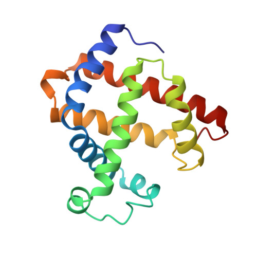
    The structure of the ferrous nitric oxide form of native sperm whale myoglobin has been determined by X-ray crystallography to 1.7 angstroms resolution. The nitric oxide ligand is bent with respect to the heme plane: the Fe-N-O angle is 112 degrees. This angle is smaller than those observed in model compounds and in lupin leghemoglobin. The exact angle appears to be influenced by the strength of the proximal bond and hydrogen bonding interactions between the distal histidine and the bound ligand. Specifically, the N(epsilon) atom of histidine64 is located 2.8 angstroms away from the nitrogen atom of the bound ligand, implying electrostatic stabilization of the FeNO complex. This interpretation is supported by mutagenesis studies. When histidine64 is replaced with apolar amino acids, the rate of nitric oxide dissociation from myoglobin increases tenfold.


  • Organizational Affiliation
    • Department of Biochemistry and Cell Biology, Rice University, Houston, Texas 77005-1892, USA.

Macromolecule Content 

  • Total Structure Weight: 18.14 kDa 
  • Atom Count: 1,416 
  • Modeled Residue Count: 154 
  • Deposited Residue Count: 154 
  • Unique protein chains: 1

Macromolecules

Find similar proteins by:|  3D Structure
Entity ID: 1
MoleculeChains  Sequence LengthOrganismDetailsImage
MYOGLOBIN154Physeter macrocephalusMutation(s): 2 
EC: 1.11.1 (UniProt), 1.7 (UniProt)
UniProt
Find proteins for P02185 (Physeter macrocephalus)
Explore P02185 
Go to UniProtKB:  P02185
Entity Groups
Sequence Clusters30% Identity50% Identity70% Identity90% Identity95% Identity100% Identity
UniProt GroupP02185
Sequence Annotations
Expand
Reference Sequence

Experimental Data & Validation

Experimental Data

  • Method: X-RAY DIFFRACTION
  • Resolution: 1.90 Å
  • R-Value Free:  0.206 (Depositor) 
  • R-Value Work:  0.142 (DCC) 
  • R-Value Observed: 0.153 (Depositor) 
Space Group: P 6
Unit Cell:
Length ( Å )Angle ( ˚ )
a = 91.506α = 90
b = 91.506β = 90
c = 45.853γ = 120
Software Package:
Software NamePurpose
SHELXL-96model building
X-PLORmodel building
SHELXL-96refinement
X-PLORrefinement
XDSdata reduction
XSCALEdata scaling
SHELXL-96phasing
X-PLORphasing

Structure Validation

View Full Validation Report


Ligand Structure Quality Assessment 


Entry History 

Deposition Data

Revision History  (Full details and data files)

  • Version 1.0: 1998-05-27
    Type: Initial release
  • Version 1.1: 2008-03-24
    Changes: Version format compliance
  • Version 1.2: 2011-07-13
    Changes: Version format compliance
  • Version 1.3: 2021-11-03
    Changes: Database references, Derived calculations, Other
  • Version 1.4: 2023-08-09
    Changes: Refinement description
  • Version 1.5: 2024-05-22
    Changes: Data collection