Substrate Recognition and Molecular Mechanism of Fatty Acid Hydroxylation by Cytochrome P450 from Bacillus subtilis. CRYSTALLOGRAPHIC, SPECTROSCOPIC, AND MUTATIONAL STUDIES.
Lee, D.S., Yamada, A., Sugimoto, H., Matsunaga, I., Ogura, H., Ichihara, K., Adachi, S., Park, S.Y., Shiro, Y.(2003) J Biological Chem 278: 9761-9767
- PubMed: 12519760 Search on PubMed
- DOI: https://doi.org/10.1074/jbc.M211575200
- Primary Citation Related Structures:
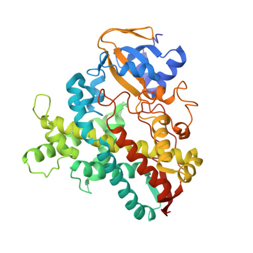
1IZO - PubMed Abstract:
Cytochrome P450 isolated from Bacillus subtilis (P450(BSbeta); molecular mass, 48 kDa) catalyzes the hydroxylation of a long-chain fatty acid (e.g. myristic acid) at the alpha- and beta-positions using hydrogen peroxide as an oxidant. We report here on the crystal structure of ferric P450(BSbeta) in the substrate-bound form, determined at a resolution of 2.1 A. P450(BSbeta) exhibits a typical P450 fold. The substrate binds to a specific channel in the enzyme and is stabilized through hydrophobic interactions of its alkyl side chain with some hydrophobic residues on the enzyme as well as by electrostatic interaction of its terminal carboxylate with the Arg(242) guanidium group. These interactions are responsible for the site specificity of the hydroxylation site in which the alpha- and beta-positions of the fatty acid come into close proximity to the heme iron sixth site. The fatty acid carboxylate group interacts with Arg(242) in the same fashion as has been reported for the active site of chloroperoxidase, His(105)-Glu(183), which is an acid-base catalyst in the peroxidation reactions. On the basis of these observations, a possible mechanism for the hydroxylation reaction catalyzed by P450(BSbeta) is proposed in which the carboxylate of the bound-substrate fatty acid assists in the cleavage of the peroxide O-O bond.
- RIKEN Harima Institute/SPring-8, 1-1-1 Kouto, Mikazuki-cho, Sayo, Hyogo 679-5148, Japan.
Organizational Affiliation: