1IGI | pdb_00001igi

26-10 FAB:DIGOXIN COMPLEX-AFFINITY AND SPECIFICITY DUE TO SURFACE COMPLEMENTARITY


Experimental Data Snapshot

  • Method: X-RAY DIFFRACTION
  • Resolution: 2.70 Å
  • R-Value Work: 
    0.177 (Depositor), 0.173 (DCC) 

wwPDB Validation 3D Report Full Report

Validation slider image for 1IGI

This is version 1.4 of the entry. See complete history

Literature

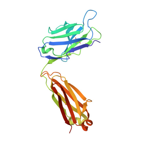
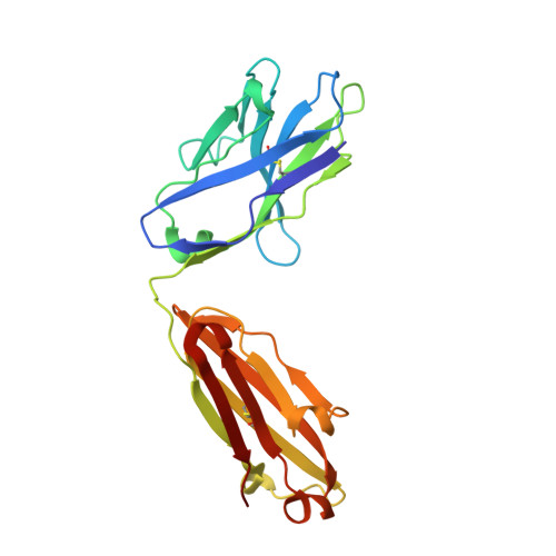
26-10 Fab-digoxin complex: affinity and specificity due to surface complementarity.

Jeffrey, P.D.Strong, R.K.Sieker, L.C.Chang, C.Y.Campbell, R.L.Petsko, G.A.Haber, E.Margolies, M.N.Sheriff, S.

(1993) Proc Natl Acad Sci U S A 90: 10310-10314

  • DOI: https://doi.org/10.1073/pnas.90.21.10310
  • Primary Citation Related Structures: 
    1IGI, 1IGJ

  • PubMed Abstract: 

    We have determined the three-dimensional structures of the antigen-binding fragment of the anti-digoxin monoclonal antibody 26-10 in the uncomplexed state at 2.7 A resolution and as a complex with digoxin at 2.5 A resolution. Neither the antibody nor digoxin undergoes any significant conformational changes upon forming the complex. Digoxin interacts primarily with the antibody heavy chain and is oriented such that the carbohydrate groups are exposed to solvent and the lactone ring is buried in a deep pocket at the bottom of the combining site. Despite extensive interactions between antibody and antigen, no hydrogen bonds or salt links are formed between 26-10 and digoxin. Thus the 26-10-digoxin complex is unique among the known three-dimensional structures of antibody-antigen complexes in that specificity and high affinity arise primarily from shape complementarity.


  • Organizational Affiliation
    • Bristol-Myers Squibb Pharmaceutical Research Institute, Princeton, NJ 08543-4000.

Macromolecule Content 

  • Total Structure Weight: 47.36 kDa 
  • Atom Count: 3,298 
  • Modeled Residue Count: 435 
  • Deposited Residue Count: 437 
  • Unique protein chains: 2

Macromolecules

Find similar proteins by:|  3D Structure
Entity ID: 1
MoleculeChains  Sequence LengthOrganismDetailsImage
IGG2A-KAPPA 26-10 FAB (LIGHT CHAIN)A [auth L]219Mus musculusMutation(s): 0 
Find similar proteins by:|  3D Structure
Entity ID: 2
MoleculeChains  Sequence LengthOrganismDetailsImage
IGG2A-KAPPA 26-10 FAB (HEAVY CHAIN)B [auth H]218Mus musculusMutation(s): 0 

Experimental Data & Validation

Experimental Data

  • Method: X-RAY DIFFRACTION
  • Resolution: 2.70 Å
  • R-Value Work:  0.177 (Depositor), 0.173 (DCC) 
Space Group: P 1 21 1
Unit Cell:
Length ( Å )Angle ( ˚ )
a = 43.78α = 90
b = 89.25β = 95.94
c = 59.24γ = 90
Software Package:
Software NamePurpose
X-PLORrefinement

Structure Validation

View Full Validation Report


Entry History 

Deposition Data

Revision History  (Full details and data files)

  • Version 1.0: 1993-04-15
    Type: Initial release
  • Version 1.1: 2008-03-24
    Changes: Version format compliance
  • Version 1.2: 2011-07-13
    Changes: Version format compliance
  • Version 1.3: 2024-06-05
    Changes: Data collection, Database references, Derived calculations, Other
  • Version 1.4: 2024-11-06
    Changes: Structure summary