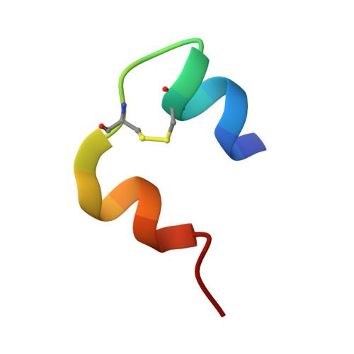
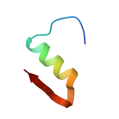
1HTV

CRYSTAL STRUCTURE OF DESTRIPEPTIDE (B28-B30) INSULIN


Experimental Data Snapshot

  • Method: X-RAY DIFFRACTION
  • Resolution: 1.90 Å
  • R-Value Free: 0.240 
  • R-Value Work: 0.196 
  • R-Value Observed: 0.196 

Starting Model: experimental
View more details

wwPDB Validation   3D Report Full Report


This is version 1.3 of the entry. See complete history


Literature

Crystal structure of destripeptide (B28-B30) insulin: implications for insulin dissociation.

Ye, J.Chang, W.Liang, D.

(2001) Biochim Biophys Acta 1547: 18-25

  • DOI: https://doi.org/10.1016/s0167-4838(01)00160-1
  • Primary Citation of Related Structures:  
    1HTV

  • PubMed Abstract: 

    Destripeptide (B28-B30) insulin (DTRI) is an insulin analogue that has much weaker association ability than native insulin but keeps most of its biological activity. It can be crystallized from a solution containing zinc ions at near-neutral pH. Its crystal structure has been determined by molecular replacement and refined at 1.9 A resolution. DTRI in the crystal exists as a loose hexamer compared with 2Zn insulin. The hexamer only contains one zinc ion that coordinates to the B10 His residues of three monomers. Although residues B28-B30 are located in the monomer-monomer interface within a dimer, the removal of them can simultaneously weaken both the interactions between monomers within the dimer and the interactions between dimers. Because the B-chain C-terminus of insulin is very flexible, we take the DTRI hexamer as a transition state in the native insulin dissociation process and suggest a possible dissociation process of the insulin hexamer based on the DTRI structure.


  • Organizational Affiliation

    National Laboratory of Biomacromolecules, Institute of Biophysics, Chinese Academy of Sciences, 15 Datun Road, Chaoyang District, 100101, Beijing, PR China.


Macromolecules

Find similar proteins by:  Sequence   |   3D Structure  

Entity ID: 1
MoleculeChains Sequence LengthOrganismDetailsImage
INSULIN
A, C, E, G, I
A, C, E, G, I, K
21Homo sapiensMutation(s): 0 
UniProt & NIH Common Fund Data Resources
Find proteins for P01308 (Homo sapiens)
Explore P01308 
Go to UniProtKB:  P01308
PHAROS:  P01308
GTEx:  ENSG00000254647 
Entity Groups  
Sequence Clusters30% Identity50% Identity70% Identity90% Identity95% Identity100% Identity
UniProt GroupP01308
Sequence Annotations
Expand
  • Reference Sequence
Find similar proteins by:  (by identity cutoff)  |  3D Structure
Entity ID: 2
MoleculeChains Sequence LengthOrganismDetailsImage
INSULIN
B, D, F, H, J
B, D, F, H, J, L
27Homo sapiensMutation(s): 0 
UniProt & NIH Common Fund Data Resources
Find proteins for P01308 (Homo sapiens)
Explore P01308 
Go to UniProtKB:  P01308
PHAROS:  P01308
GTEx:  ENSG00000254647 
Entity Groups  
Sequence Clusters30% Identity50% Identity70% Identity90% Identity95% Identity100% Identity
UniProt GroupP01308
Sequence Annotations
Expand
  • Reference Sequence
Small Molecules
Ligands 1 Unique
IDChains Name / Formula / InChI Key2D Diagram3D Interactions
ZN
Query on ZN

Download Ideal Coordinates CCD File 
M [auth B]ZINC ION
Zn
PTFCDOFLOPIGGS-UHFFFAOYSA-N
Experimental Data & Validation

Experimental Data

  • Method: X-RAY DIFFRACTION
  • Resolution: 1.90 Å
  • R-Value Free: 0.240 
  • R-Value Work: 0.196 
  • R-Value Observed: 0.196 
  • Space Group: P 21 21 21
Unit Cell:
Length ( Å )Angle ( ˚ )
a = 49.81α = 90
b = 51.55β = 90
c = 100.6γ = 90
Software Package:
Software NamePurpose
DENZOdata reduction
SCALEPACKdata scaling
AMoREphasing
X-PLORrefinement

Structure Validation

View Full Validation Report


Entry History 

Deposition Data

Revision History  (Full details and data files)

  • Version 1.0: 2001-05-23
    Type: Initial release
  • Version 1.1: 2008-04-27
    Changes: Version format compliance
  • Version 1.2: 2011-07-13
    Changes: Version format compliance
  • Version 1.3: 2024-04-03
    Changes: Data collection, Database references, Derived calculations, Refinement description