1HTQ | pdb_00001htq

Multicopy crystallographic structure of a relaxed glutamine synthetase from Mycobacterium tuberculosis


Experimental Data Snapshot

  • Method: X-RAY DIFFRACTION
  • Resolution: 2.40 Å
  • R-Value Free: 
    0.223 (Depositor) 
  • R-Value Work: 
    0.204 (Depositor), 0.219 (DCC) 
  • R-Value Observed: 
    0.205 (Depositor) 

Starting Model: experimental
View more details

wwPDB Validation 3D Report Full Report

Validation slider image for 1HTQ

Ligand Structure Quality Assessment 


This is version 1.5 of the entry. See complete history

Literature

Multicopy crystallographic refinement of a relaxed glutamine synthetase from Mycobacterium tuberculosis highlights flexible loops in the enzymatic mechanism and its regulation.

Gill, H.S.Pfluegl, G.M.Eisenberg, D.

(2002) Biochemistry 41: 9863-9872

  • DOI: https://doi.org/10.1021/bi020254s
  • Primary Citation Related Structures: 
    1HTO, 1HTQ

  • PubMed Abstract: 

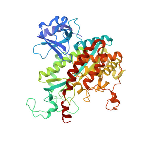
    The crystal structure of glutamine synthetase (GS) from Mycobacterium tuberculosis determined at 2.4 A resolution reveals citrate and AMP bound in the active site. The structure was refined with strict 24-fold noncrystallographic symmetry (NCS) constraints and has an R-factor of 22.7% and an R-free of 25.5%. Multicopy refinement using 10 atomic models and strict 24-fold NCS constraints further reduced the R-factor to 20.4% and the R-free to 23.2%. The multicopy model demonstrates the range of atomic displacements of catalytic and regulatory loops in glutamine synthesis, simulating loop motions. A comparison with loop positions in substrate complexes of GS from Salmonella typhimurium shows that the Asp50 and Glu327 loops close over the active site during catalysis. These loop closures are preceded by a conformational change of the Glu209 beta-strand upon metal ion or ATP binding that converts the enzyme from a relaxed to a taut state. We propose a model of the GS regulatory mechanism based on the loop motions in which adenylylation of the Tyr397 loop reverses the effect of metal ion binding, and regulates intermediate formation by preventing closure of the Glu327 loop.


  • Organizational Affiliation
    • Howard Hughes Medical Institute, UCLA-Department of Energy Laboratory of Structural Biology and Molecular Medicine, Department of Chemistry and Biochemistry, Box 951570, University of California, Los Angeles, CA 90095-1570, USA.

Macromolecule Content 

  • Total Structure Weight: 1,298.18 kDa 
  • Atom Count: 97,872 
  • Modeled Residue Count: 11,448 
  • Deposited Residue Count: 11,448 
  • Unique protein chains: 1

Macromolecules

Find similar proteins by:|  3D Structure
Entity ID: 1
MoleculeChains  Sequence LengthOrganismDetailsImage
glutamine synthetase
A, B, C, D, E
A, B, C, D, E, F, G, H, I, J, K, L, M, N, O, P, Q, R, S, T, U, V, W, X
477Mycobacterium tuberculosisMutation(s): 0 
EC: 6.3.1.2
UniProt
Find proteins for P9WN39 (Mycobacterium tuberculosis (strain ATCC 25618 / H37Rv))
Explore P9WN39 
Go to UniProtKB:  P9WN39
Entity Groups
Sequence Clusters30% Identity50% Identity70% Identity90% Identity95% Identity100% Identity
UniProt GroupP9WN39
Sequence Annotations
Expand
Reference Sequence

Small Molecules

Ligands 3 Unique
IDChains Name / Formula / InChI Key2D Diagram3D Interactions
AMP

Query on AMP


Download:Ideal Coordinates CCD File
AB [auth J]
BC [auth S]
CA [auth B]
DB [auth K]
EC [auth T]
AB [auth J],
BC [auth S],
CA [auth B],
DB [auth K],
EC [auth T],
FA [auth C],
GB [auth L],
HC [auth U],
IA [auth D],
JB [auth M],
KC [auth V],
LA [auth E],
MB [auth N],
NC [auth W],
OA [auth F],
PB [auth O],
QC [auth X],
RA [auth G],
SB [auth P],
UA [auth H],
VB [auth Q],
XA [auth I],
YB [auth R],
Z [auth A]
ADENOSINE MONOPHOSPHATE
C10 H14 N5 O7 P
UDMBCSSLTHHNCD-KQYNXXCUSA-N
CIT

Query on CIT


Download:Ideal Coordinates CCD File
AA [auth A]
BB [auth J]
CC [auth S]
DA [auth B]
EB [auth K]
AA [auth A],
BB [auth J],
CC [auth S],
DA [auth B],
EB [auth K],
FC [auth T],
GA [auth C],
HB [auth L],
IC [auth U],
JA [auth D],
KB [auth M],
LC [auth V],
MA [auth E],
NB [auth N],
OC [auth W],
PA [auth F],
QB [auth O],
RC [auth X],
SA [auth G],
TB [auth P],
VA [auth H],
WB [auth Q],
YA [auth I],
ZB [auth R]
CITRIC ACID
C6 H8 O7
KRKNYBCHXYNGOX-UHFFFAOYSA-N
MN

Query on MN


Download:Ideal Coordinates CCD File
AC [auth S]
BA [auth B]
CB [auth K]
DC [auth T]
EA [auth C]
AC [auth S],
BA [auth B],
CB [auth K],
DC [auth T],
EA [auth C],
FB [auth L],
GC [auth U],
HA [auth D],
IB [auth M],
JC [auth V],
KA [auth E],
LB [auth N],
MC [auth W],
NA [auth F],
OB [auth O],
PC [auth X],
QA [auth G],
RB [auth P],
TA [auth H],
UB [auth Q],
WA [auth I],
XB [auth R],
Y [auth A],
ZA [auth J]
MANGANESE (II) ION
Mn
WAEMQWOKJMHJLA-UHFFFAOYSA-N

Experimental Data & Validation

Experimental Data

  • Method: X-RAY DIFFRACTION
  • Resolution: 2.40 Å
  • R-Value Free:  0.223 (Depositor) 
  • R-Value Work:  0.204 (Depositor), 0.219 (DCC) 
  • R-Value Observed: 0.205 (Depositor) 
Space Group: P 21 21 21
Unit Cell:
Length ( Å )Angle ( ˚ )
a = 207.72α = 90
b = 257.69β = 90
c = 274.5γ = 90
Software Package:
Software NamePurpose
X-PLORmodel building
X-PLORrefinement
MOSFLMdata reduction
CCP4data scaling
ROTAVATAdata scaling
X-PLORphasing

Structure Validation

View Full Validation Report


Ligand Structure Quality Assessment 


Entry History 

Deposition Data

Revision History  (Full details and data files)

  • Version 1.0: 2002-07-24
    Type: Initial release
  • Version 1.1: 2008-04-27
    Changes: Version format compliance
  • Version 1.2: 2011-07-13
    Changes: Version format compliance
  • Version 1.3: 2015-09-09
    Changes: Version format compliance
  • Version 1.4: 2017-10-04
    Changes: Refinement description
  • Version 1.5: 2023-08-09
    Changes: Data collection, Database references, Derived calculations, Refinement description