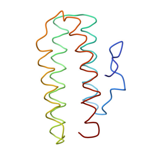
Structure of trimeric haemerythrin.
Smith, J.L., Hendrickson, W.A., Addison, A.W.(1983) Nature 303: 86-88
- PubMed: 6843663 Search on PubMed
- DOI: https://doi.org/10.1038/303086a0
- Primary Citation Related Structures:
1HR3 - PubMed Abstract:
Several simplifying structural principles have been developed from the considerable data contained in the three-dimensional structures of proteins determined in the past two decades. One of these is based on the observation that particular folding motifs often occur in a variety of structural and functional settings. The compact bundle of four antiparallel alpha-helices, first seen in the structure of myohaemerythrin, is an example. Several non-haemerythrin proteins have since been found to have the same folding pattern, and haemerythrins themselves exist in a wide variety of quaternary arrangements. The unusual ability of the haemerythrin fold to associate as dimers, trimers, tetramers, octamers or higher aggregates provides an opportunity for examining structural diversity in subunit association. We have used X-ray crystallography to study the subunit structure of trimeric haemerythrin from a Siphonosoma species. We report here that the pattern of intersubunit helix-helix interactions differs from the most common mode of association of other helix-bundle proteins. In a novel approach to structure analysis at low resolution, experimental phases for the structure determination were based on anomalous scattering from the iron atoms native to haemerythrin, using the new resolved-anomalous phasing procedure.