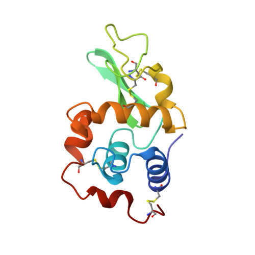
Structural and thermodynamic analysis of compensating mutations within the core of chicken egg white lysozyme.
Wilson, K.P., Malcolm, B.A., Matthews, B.W.(1992) J Biological Chem 267: 10842-10849
- PubMed: 1587860 Search on PubMed
- Primary Citation Related Structures:
1HEL, 1HEM, 1HEN, 1HEO, 1HEP, 1HEQ, 1HER - PubMed Abstract:
High resolution crystal structures have been determined for six chicken-type lysozymes that were constructed to investigate putative intermediates in the evolution of the lysozymes of modern game birds (Malcolm, B. A., Wilson, K. P., Matthews, B. W., Kirsch, J. F., and Wilson, A. C. (1990) Nature 345, 86-89). The amino acid replacements include Thr-40----Ser, Ile-55----Val, and Ser-91----Thr, as well as combinations of these substitutions. Residues 40, 55, and 91 are buried within the core of chicken lysozyme. The replacements therefore involve the insertion and/or removal of methyl groups from the protein interior. The mutant proteins have normal activities, and their thermal stabilities span a range of 7 degrees C, with some variants more stable and some less stable than the naturally occurring forms. Comparison of the crystal structures shows the overall structures to be very similar, but there are differences in the packing of side chains in the region of the replacements. The x-ray coordinates were used to evaluate the repacking of side chains in the protein interior and to attempt to evaluate the contributions of the different energetic interactions toward the overall stability of each variant. The results illustrate how proteins can compensate for potentially destabilizing substitutions in different ways and underscore the importance of high resolution structural data if changes in protein thermostability due to changes in protein sequence are to be understood. The findings also suggest that protein stability can be increased by mutations that lower strain in the protein interior while maintaining total buried hydrophobic surface area.
- Institute of Molecular Biology, Howard Hughes Medical Institute, Eugene, Oregon.
Organizational Affiliation: