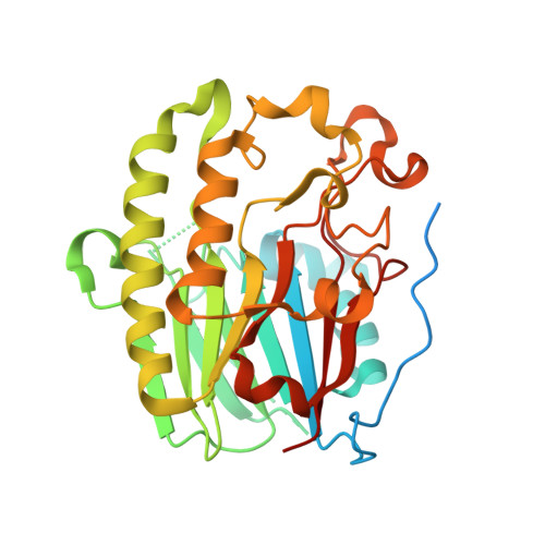
1HD7

A Second Divalent Metal Ion in the Active Site of a New Crystal Form of Human Apurinic/Apyridinimic Endonuclease, Ape1, and its Implications for the Catalytic Mechanism


Experimental Data Snapshot

  • Method: X-RAY DIFFRACTION
  • Resolution: 1.95 Å
  • R-Value Free: 0.255 
  • R-Value Work: 0.205 

Starting Model: other
View more details

wwPDB Validation   3D Report Full Report


This is version 1.3 of the entry. See complete history


Literature

Two Divalent Metal Ions in the Active Site of a New Crystal Form of Human Apurinic/Apyrimidinic Endonuclease, Ape1: Implications for the Catalytic Mechanism

Beernink, P.T.Segelke, B.W.Hadi, M.Z.Erzberger, J.P.Wilson III, D.M.Rupp, B.

(2001) J Mol Biol 307: 1023

  • DOI: https://doi.org/10.1006/jmbi.2001.4529
  • Primary Citation of Related Structures:  
    1E9N, 1HD7

  • PubMed Abstract: 

    The major human abasic endonuclease, Ape1, is an essential DNA repair enzyme that initiates the removal of apurinic/apyrimidinic sites from DNA, excises 3' replication-blocking moieties, and modulates the DNA binding activity of several transcriptional regulators. We have determined the X-ray structure of the full-length human Ape1 enzyme in two new crystal forms, one at neutral and one at acidic pH. The new structures are generally similar to the previously determined structure of a truncated Ape1 protein, but differ in the conformation of several loop regions and in spans of residues with weak electron density. While only one active-site metal ion is present in the structure determined at low pH, the structure determined from a crystal grown at the pH optimum of Ape1 nuclease activity, pH 7.5, has two metal ions bound 5 A apart in the active site. Enzyme kinetic data indicate that at least two metal-binding sites are functionally important, since Ca(2+) exhibits complex stimulatory and inhibitory effects on the Mg(2+)-dependent catalysis of Ape1, even though Ca(2+) itself does not serve as a cofactor. In conjunction, the structural and kinetic data suggest that Ape1 catalyzes hydrolysis of the DNA backbone through a two metal ion-mediated mechanism.


  • Organizational Affiliation

    Molecular and Structural Biology Division, Biology and Biotechnology Research Program, Lawrence Livermore National Laboratory, Livermore, CA, 94550, USA.


Macromolecules
Find similar proteins by:  (by identity cutoff)  |  3D Structure
Entity ID: 1
MoleculeChains Sequence LengthOrganismDetailsImage
DNA-(APURINIC OR APYRIMIDINIC SITE) LYASE318Homo sapiensMutation(s): 0 
EC: 4.2.99.18
UniProt & NIH Common Fund Data Resources
Find proteins for P27695 (Homo sapiens)
Explore P27695 
Go to UniProtKB:  P27695
PHAROS:  P27695
GTEx:  ENSG00000100823 
Entity Groups  
Sequence Clusters30% Identity50% Identity70% Identity90% Identity95% Identity100% Identity
UniProt GroupP27695
Sequence Annotations
Expand
  • Reference Sequence
Small Molecules
Ligands 1 Unique
IDChains Name / Formula / InChI Key2D Diagram3D Interactions
PB
Query on PB

Download Ideal Coordinates CCD File 
B [auth A]LEAD (II) ION
Pb
RVPVRDXYQKGNMQ-UHFFFAOYSA-N
Experimental Data & Validation

Experimental Data

  • Method: X-RAY DIFFRACTION
  • Resolution: 1.95 Å
  • R-Value Free: 0.255 
  • R-Value Work: 0.205 
  • Space Group: C 1 2 1
Unit Cell:
Length ( Å )Angle ( ˚ )
a = 128.39α = 90
b = 44.85β = 124.54
c = 78.14γ = 90
Software Package:
Software NamePurpose
REFMACrefinement
MOSFLMdata reduction
SCALAdata scaling
EPMRphasing

Structure Validation

View Full Validation Report


Entry History 

Deposition Data

Revision History  (Full details and data files)

  • Version 1.0: 2001-02-16
    Type: Initial release
  • Version 1.1: 2011-05-08
    Changes: Version format compliance
  • Version 1.2: 2011-07-13
    Changes: Version format compliance
  • Version 1.3: 2024-05-01
    Changes: Data collection, Database references, Derived calculations, Other, Refinement description