The Er Protein Folding Sensor Udp-Glucose Glycoprotein:Glucosyltransferase Modifies Substrates Distant to Local Changes in Glycoprotein Conformation.
Taylor, S.C., Ferguson, A.D., Bergeron, J.J.M., Thomas, D.Y.(2004) Nat Struct Mol Biol 11: 128
- PubMed: 14730348 Search on PubMed
- DOI: https://doi.org/10.1038/nsmb715
- Primary Citation Related Structures:
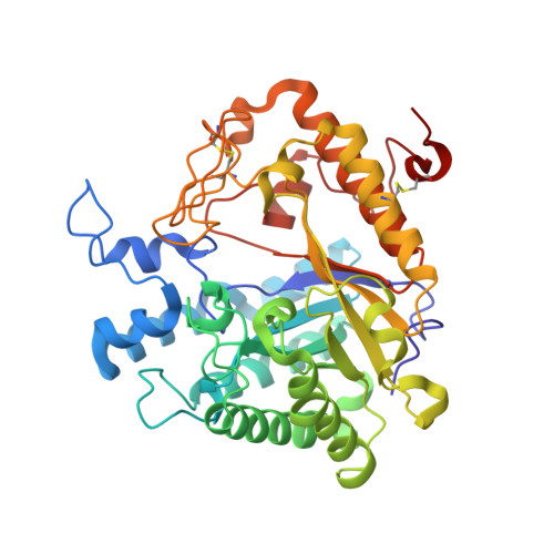
1H4P - PubMed Abstract:
We present in vitro data that explain the recognition mechanism of misfolded glycoproteins by UDP-glucose glycoprotein-glucosyltransferase (UGGT). The glycoprotein exo-(1,3)-beta-glucanase (beta-Glc) bearing two glycans unfolds in a pH-dependent manner to become a misfolded substrate for UGGT. In the crystal structure of this glycoprotein, the local hydrophobicity surrounding each glycosylation site coincides with the differential recognition of N-linked glycans by UGGT. We introduced a single F280S point mutation, producing a beta-Glc protein with full enzymatic activity that was both recognized as misfolded and monoglucosylated by UGGT. Contrary to current views, these data show that UGGT can modify N-linked glycans positioned at least 40 A from localized regions of disorder and sense subtle conformational changes within structurally compact, enzymatically active glycoprotein substrates.
- Biochemistry Department, Faculty of Medicine, McGill University, McIntyre Medical Sciences Building, 3655 Boulevard Sir William Osler, Montreal, Quebec, Canada, H3G 1Y6.
Organizational Affiliation: