Electrostatic Effects and Binding Determinants in the Catalysis of Prolyl Oligopeptidase: Site Specific Mutagenesis at the Oxyanion Binding Site
Szeltner, Z., Rea, D., Renner, V., Fulop, V., Polgar, L.(2002) J Biological Chem 277: 42613
- PubMed: 12202494 Search on PubMed
- DOI: https://doi.org/10.1074/jbc.M208043200
- Primary Citation Related Structures:
1H2W, 1H2X, 1H2Y, 1H2Z - PubMed Abstract:
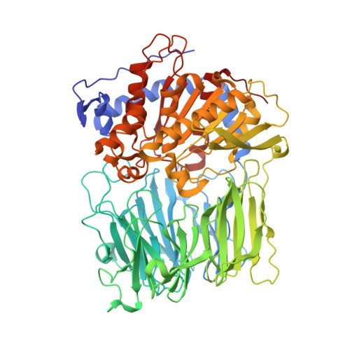
Prolyl oligopeptidase, a member of a new family of serine peptidases, plays an important role in memory disorders. Earlier x-ray crystallographic investigations indicated that stabilization of the tetrahedral transition state of the reaction involved hydrogen bond formation between the oxyanion of the tetrahedral intermediate and the OH group of Tyr(473). The contribution of the OH group was tested with the Y473F variant using various substrates. The charged succinyl-Gly-Pro-4-nitroanilide was hydrolyzed with a much lower k(cat)/K(m) compared with the neutral benzyloxycarbonyl-G1y-Pro-2-naphthylamide, although the binding modes of the two substrates were similar, as shown by x-ray crystallography. This suggested that electrostatic interactions between Arg(643) and the succinyl group competed with the productive binding mechanism. Unlike most enzyme reactions, catalysis by the wild-type enzyme exhibited positive activation entropy. In contrast, the activation entropy for the Y473F variant was negative, suggesting that the tyrosine OH group is involved in stabilizing both the transition state and the water shell at the active site. Importantly, Tyr(473) is also implicated in the formation of the enzyme-substrate complex. The nonlinear Arrhenius plot suggested a greater significance of the oxyanion binding site at physiological temperature. The results indicated that Tyr(473) was more needed at high pH, at high temperature, and with charged substrates exhibiting "internally competitive inhibition."
- Institute of Enzymology, Biological Research Center, Hungarian Academy of Sciences, H-1518 Budapest, Hungary.
Organizational Affiliation: