1H15

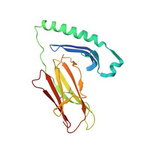
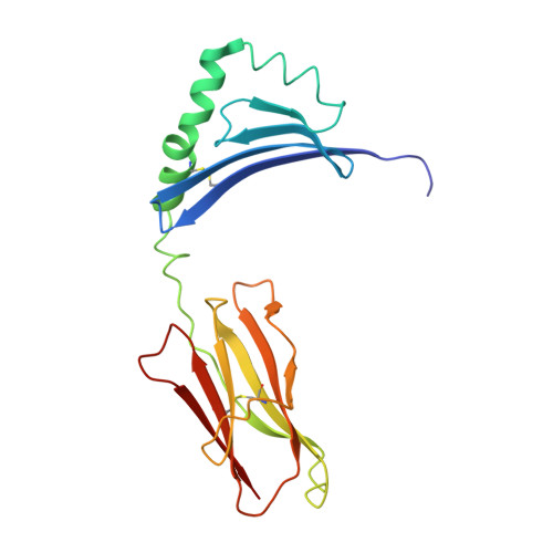
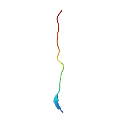
X-ray crystal structure of HLA-DRA1*0101/DRB5*0101 complexed with a peptide from Epstein Barr Virus DNA polymerase


Experimental Data Snapshot

  • Method: X-RAY DIFFRACTION
  • Resolution: 3.10 Å
  • R-Value Free: 0.310 
  • R-Value Work: 0.256 
  • R-Value Observed: 0.256 

Starting Model: experimental
View more details

wwPDB Validation   3D Report Full Report


Ligand Structure Quality Assessment 


This is version 2.1 of the entry. See complete history


Literature

A Functional and Structural Basis for Tcr Cross-Reactivity in Multiple Sclerosis

Lang, H.Jacobsen, H.Ikemizu, S.Andersson, C.Harlos, K.Madsen, L.Hjorth, P.Sondergaard, L.Svejgaard, A.Wucherpfennig, K.Stuart, D.I.Bell, J.I.Jones, E.Y.Fugger, L.

(2002) Nat Immunol 3: 940

  • DOI: https://doi.org/10.1038/ni835
  • Primary Citation of Related Structures:  
    1H15

  • PubMed Abstract: 

    The multiple sclerosis (MS)-associated HLA major histocompatibility complex (MHC) class II alleles DRB1*1501, DRB5*0101 and DQB1*0602 are in strong linkage disequilibrium, making it difficult to determine which is the principal MS risk gene. Here we show that together the DRB1 and DRB5 loci may influence susceptibility to MS. We demonstrate that a T cell receptor (TCR) from an MS patient recognized both a DRB1*1501-restricted myelin basic protein (MBP) and DRB5*0101-restricted Epstein-Barr virus (EBV) peptide. Crystal structure determination of the DRB5*0101-EBV peptide complex revealed a marked degree of structural equivalence to the DRB1*1501-MBP peptide complex at the surface presented for TCR recognition. This provides structural evidence for molecular mimicry involving HLA molecules. The structural details suggest an explanation for the preponderance of MHC class II associations in HLA-associated diseases.


  • Organizational Affiliation

    Nuffield Department of Clinical Medicine, John Radcliffe Hospital, Headington, Oxford OX3 9DU, UK.


Macromolecules
Find similar proteins by:  (by identity cutoff)  |  3D Structure
Entity ID: 1
MoleculeChains Sequence LengthOrganismDetailsImage
HLA CLASS II HISTOCOMPATIBILITY ANTIGEN, DR ALPHA CHAIN
A, D
182Homo sapiensMutation(s): 0 
UniProt & NIH Common Fund Data Resources
Find proteins for P01903 (Homo sapiens)
Explore P01903 
Go to UniProtKB:  P01903
PHAROS:  P01903
GTEx:  ENSG00000204287 
Entity Groups  
Sequence Clusters30% Identity50% Identity70% Identity90% Identity95% Identity100% Identity
UniProt GroupP01903
Glycosylation
Glycosylation Sites: 2Go to GlyGen: P01903-1
Sequence Annotations
Expand
  • Reference Sequence
Find similar proteins by:  (by identity cutoff)  |  3D Structure
Entity ID: 2
MoleculeChains Sequence LengthOrganismDetailsImage
HLA CLASS II HISTOCOMPATIBILITY ANTIGEN, DR BETA 1 CHAIN
B, E
190Homo sapiensMutation(s): 0 
UniProt & NIH Common Fund Data Resources
Find proteins for Q30154 (Homo sapiens)
Explore Q30154 
Go to UniProtKB:  Q30154
PHAROS:  Q30154
GTEx:  ENSG00000198502 
Entity Groups  
Sequence Clusters30% Identity50% Identity70% Identity90% Identity95% Identity100% Identity
UniProt GroupQ30154
Sequence Annotations
Expand
  • Reference Sequence

Find similar proteins by:  Sequence   |   3D Structure  

Entity ID: 3
MoleculeChains Sequence LengthOrganismDetailsImage
DNA POLYMERASE
C, F
14human gammaherpesvirus 4Mutation(s): 0 
EC: 2.7.7.7
UniProt
Find proteins for P03198 (Epstein-Barr virus (strain B95-8))
Explore P03198 
Go to UniProtKB:  P03198
Entity Groups  
Sequence Clusters30% Identity50% Identity70% Identity90% Identity95% Identity100% Identity
UniProt GroupP03198
Sequence Annotations
Expand
  • Reference Sequence
Oligosaccharides

Help

Entity ID: 4
MoleculeChains Length2D Diagram Glycosylation3D Interactions
2-acetamido-2-deoxy-beta-D-glucopyranose-(1-4)-2-acetamido-2-deoxy-beta-D-glucopyranose
G
2N-Glycosylation
Glycosylation Resources
GlyTouCan:  G42666HT
GlyCosmos:  G42666HT
GlyGen:  G42666HT
Experimental Data & Validation

Experimental Data

  • Method: X-RAY DIFFRACTION
  • Resolution: 3.10 Å
  • R-Value Free: 0.310 
  • R-Value Work: 0.256 
  • R-Value Observed: 0.256 
  • Space Group: P 65
Unit Cell:
Length ( Å )Angle ( ˚ )
a = 179.244α = 90
b = 179.244β = 90
c = 92.884γ = 120
Software Package:
Software NamePurpose
CNSrefinement
DENZOdata reduction
SCALEPACKdata scaling
AMoREphasing

Structure Validation

View Full Validation Report


Ligand Structure Quality Assessment 


Entry History 

Revision History  (Full details and data files)

  • Version 1.0: 2002-10-03
    Type: Initial release
  • Version 1.1: 2011-09-28
    Changes: Advisory, Atomic model, Derived calculations, Non-polymer description, Other, Refinement description, Version format compliance
  • Version 1.2: 2012-06-27
    Changes: Other
  • Version 2.0: 2020-07-29
    Type: Remediation
    Reason: Carbohydrate remediation
    Changes: Advisory, Atomic model, Data collection, Derived calculations, Other, Structure summary
  • Version 2.1: 2023-12-13
    Changes: Data collection, Database references, Refinement description, Structure summary