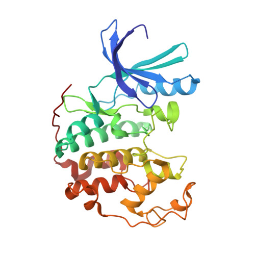
1H0V

Human cyclin dependent protein kinase 2 in complex with the inhibitor 2-Amino-6-[(R)-pyrrolidino-5'-yl]methoxypurine


Experimental Data Snapshot

  • Method: X-RAY DIFFRACTION
  • Resolution: 1.90 Å
  • R-Value Free: 0.234 
  • R-Value Work: 0.154 
  • R-Value Observed: 0.158 

Starting Model: experimental
View more details

wwPDB Validation   3D Report Full Report


Ligand Structure Quality Assessment 


This is version 1.4 of the entry. See complete history


Literature

Probing the ATP Ribose-Binding Domain of Cyclin-Dependent Kinases 1 and 2 with O(6)-Substituted Guanine Derivatives

Gibson, A.E.Arris, C.E.Bentley, J.Boyle, F.T.Curtin, N.J.Davies, T.G.Endicott, J.A.Golding, B.T.Grant, S.Griffin, R.J.Jewsbury, P.Johnson, L.N.Mesguiche, V.Newell, D.R.Noble, M.E.M.Tucker, J.A.Whitfield, H.J.

(2002) J Med Chem 45: 3381

  • DOI: https://doi.org/10.1021/jm020056z
  • Primary Citation of Related Structures:  
    1GZ8, 1H0V, 1H0W

  • PubMed Abstract: 

    O(6)-substituted guanines are adenosine 5'-triphosphate (ATP) competitive inhibitors of CDK1/cyclin B1 and CDK2/cyclin A, the O(6) substituent occupying the kinase ribose binding site. Fifty-eight O(6)-substituted guanines were prepared to probe the ribose pocket, and the structures of four representative compounds bound to monomeric CDK2 were determined by X-ray crystallography. Optimum binding occurs with a moderately sized aliphatic O(6) substituent that packs tightly against the hydrophobic patch presented by the glycine loop, centered on Val18, an interaction promoted by the conformational restraints imposed in a cyclohexylmethyl or cyclohexenylmethyl ring. Structure-based design generated (R)-(2-amino-9H-purin-6-yloxymethyl)pyrrolidin-2-one (56), which reproduces the reported hydrogen bonds formed between ATP and Asp86 and Gln131 but failed to improve inhibitory potency. Thus, the parent compound O(6)-cyclohexylmethylguanine (NU2058, 25) is the preferred starting point for exploring other areas of the kinase active site.


  • Organizational Affiliation

    Department of Chemistry and Cancer Research Unit, University of Newcastle, Newcastle upon Tyne NE2 4HH, United Kingdom.


Macromolecules
Find similar proteins by:  (by identity cutoff)  |  3D Structure
Entity ID: 1
MoleculeChains Sequence LengthOrganismDetailsImage
CELL DIVISION PROTEIN KINASE 2298Homo sapiensMutation(s): 0 
EC: 2.7.1.37 (PDB Primary Data), 2.7.11.22 (UniProt)
UniProt & NIH Common Fund Data Resources
Find proteins for P24941 (Homo sapiens)
Explore P24941 
Go to UniProtKB:  P24941
PHAROS:  P24941
GTEx:  ENSG00000123374 
Entity Groups  
Sequence Clusters30% Identity50% Identity70% Identity90% Identity95% Identity100% Identity
UniProt GroupP24941
Sequence Annotations
Expand
  • Reference Sequence
Small Molecules
Ligands 1 Unique
IDChains Name / Formula / InChI Key2D Diagram3D Interactions
UN4
Query on UN4

Download Ideal Coordinates CCD File 
B [auth A]5-{[(2-AMINO-9H-PURIN-6-YL)OXY]METHYL}-2-PYRROLIDINONE
C10 H12 N6 O2
UYPMMHCTXQIWDX-RXMQYKEDSA-N
Experimental Data & Validation

Experimental Data

  • Method: X-RAY DIFFRACTION
  • Resolution: 1.90 Å
  • R-Value Free: 0.234 
  • R-Value Work: 0.154 
  • R-Value Observed: 0.158 
  • Space Group: P 21 21 21
Unit Cell:
Length ( Å )Angle ( ˚ )
a = 52.953α = 90
b = 70.912β = 90
c = 72.283γ = 90
Software Package:
Software NamePurpose
REFMACrefinement
DENZOdata reduction
SCALEPACKdata scaling
CCP4phasing

Structure Validation

View Full Validation Report


Ligand Structure Quality Assessment 


Entry History 

Revision History  (Full details and data files)

  • Version 1.0: 2003-06-27
    Type: Initial release
  • Version 1.1: 2011-05-08
    Changes: Version format compliance
  • Version 1.2: 2011-07-13
    Changes: Version format compliance
  • Version 1.3: 2019-03-06
    Changes: Data collection, Experimental preparation, Other
  • Version 1.4: 2023-12-13
    Changes: Data collection, Database references, Refinement description