Structural Basis of Membrane-Induced Cardiotoxin A3 Oligomerization
Forouhar, F., Huang, W.-N., Liu, J.-H., Chien, K.-Y., Wu, W.-G., Hsiao, C.-D.(2003) J Biological Chem 278: 21980
- PubMed: 12660250 Search on PubMed
- DOI: https://doi.org/10.1074/jbc.M208650200
- Primary Citation Related Structures:
1H0J - PubMed Abstract:
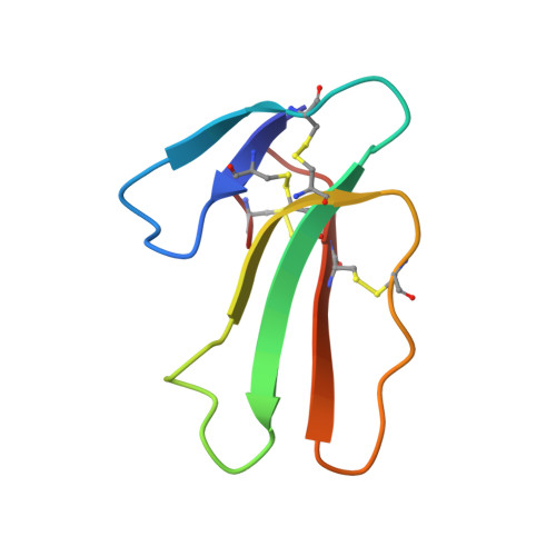
Cobra cardiotoxins (CTXs) have previously been shown to induce membrane fusion of vesicles formed by phospholipids such as cardiolipin or sphingomyelin. CTX can also form a pore in membrane bilayers containing a anionic lipid such as phosphatidylserine or phosphatidylglycerol. Herein, we show that the interaction of CTX with negatively charged lipids causes CTX dimerization, an important intermediate for the eventual oligomerization of CTX during the CTX-induced fusion and pore formation process. The structural basis of the lipid-induced oligomerization of CTX A3, a major CTX from Naja atra, is then illustrated by the crystal structure of CTX A3 in complex with SDS; SDS likely mimics anionic lipids of the membrane under micelle conditions at 1.9-A resolution. The crystal packing reveals distinct SDS-free and SDS-rich regions; in the latter two types of interconnecting CTX A3 dimers, D1 and D2, and several SDS molecules can be identified to stabilize D1 and D2 by simultaneously interacting with residues at each dimer interface. When the three CTXSDS complexes in the asymmetric unit are overlaid, the orientation of CTX A3 monomers relative to the SDS molecules in the crystal is strikingly similar to that of the toxin with respect to model membranes as determined by NMR and Fourier transform infrared methods. These results not only illustrate how lipid-induced CTX dimer formation may be transformed into oligomers either as inverted micelles of fusion intermediates or as membrane pore of anionic lipid bilayers but also underscore a potential role for SDS in x-ray diffraction study of protein-membrane interactions in the future.
- Institute of Molecular Biology, Academia Sinica, Taipei, Taiwan 115.
Organizational Affiliation: