1GWB | pdb_00001gwb

STRUCTURE OF GLYCOPROTEIN 1B


Experimental Data Snapshot

  • Method: X-RAY DIFFRACTION
  • Resolution: 2.80 Å
  • R-Value Free: 
    0.274 (Depositor), 0.266 (DCC) 
  • R-Value Work: 
    0.245 (Depositor), 0.241 (DCC) 
  • R-Value Observed: 
    0.245 (Depositor) 

wwPDB Validation 3D Report Full Report

Validation slider image for 1GWB

Ligand Structure Quality Assessment 


This is version 1.5 of the entry. See complete history

Literature

Crystal Structure of the Platelet Glycoprotein Ib-Alpha N-Terminal Domain Reveals an Unmasking Mechanism of Receptor Activation

Uff, S.Clemetson, J.M.Harrison, T.Clemetson, K.J.Emsley, J.

(2002) J Biological Chem 277: 35657

  • DOI: https://doi.org/10.1074/jbc.M205271200
  • Primary Citation Related Structures: 
    1GWB

  • PubMed Abstract: 

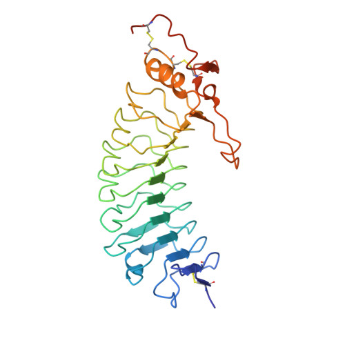
    Glycoprotein Ib (GPIb) is a platelet receptor with a critical role in mediating the arrest of platelets at sites of vascular damage. GPIb binds to the A1 domain of von Willebrand factor (vWF-A1) at high blood shear, initiating platelet adhesion and contributing to the formation of a thrombus. To investigate the molecular basis of GPIb regulation and ligand binding, we have determined the structure of the N-terminal domain of the GPIb(alpha) chain (residues 1-279). This structure is the first determined from the cell adhesion/signaling class of leucine-rich repeat (LRR) proteins and reveals the topology of the characteristic disulfide-bonded flanking regions. The fold consists of an N-terminal beta-hairpin, eight leucine-rich repeats, a disulfide-bonded loop, and a C-terminal anionic region. The structure also demonstrates a novel LRR motif in the form of an M-shaped arrangement of three tandem beta-turns. Negatively charged binding surfaces on the LRR concave face and anionic region indicate two-step binding kinetics to vWF-A1, which can be regulated by an unmasking mechanism involving conformational change of a key loop. Using molecular docking of the GPIb and vWF-A1 crystal structures, we were also able to model the GPIb.vWF-A1 complex.


  • Organizational Affiliation
    • Department of Biochemistry, University of Leicester, Leicester LE1 7RH, United Kingdom.

Macromolecule Content 

  • Total Structure Weight: 64.63 kDa 
  • Atom Count: 4,451 
  • Modeled Residue Count: 545 
  • Deposited Residue Count: 562 
  • Unique protein chains: 1

Macromolecules

Find similar proteins by:|  3D Structure
Entity ID: 1
MoleculeChains  Sequence LengthOrganismDetailsImage
PLATELET GLYCOPROTEIN IB ALPHA CHAIN
A, B
281Homo sapiensMutation(s): 0 
UniProt & NIH Common Fund Data Resources
Find proteins for P07359 (Homo sapiens)
Explore P07359 
Go to UniProtKB:  P07359
PHAROS:  P07359
GTEx:  ENSG00000185245 
Entity Groups
Sequence Clusters30% Identity50% Identity70% Identity90% Identity95% Identity100% Identity
UniProt GroupP07359
Glycosylation
Glycosylation Sites: 2Go to GlyGen: P07359-1
Sequence Annotations
Expand
Reference Sequence

Small Molecules

Ligands 4 Unique
IDChains Name / Formula / InChI Key2D Diagram3D Interactions
NAG

Query on NAG


Download:Ideal Coordinates CCD File
F [auth A],
G [auth A],
J [auth B]
2-acetamido-2-deoxy-beta-D-glucopyranose
C8 H15 N O6
OVRNDRQMDRJTHS-FMDGEEDCSA-N
PT

Query on PT


Download:Ideal Coordinates CCD File
C [auth A],
D [auth A],
H [auth B],
I [auth B]
PLATINUM (II) ION
Pt
HRGDZIGMBDGFTC-UHFFFAOYSA-N
SO4

Query on SO4


Download:Ideal Coordinates CCD File
K [auth B]SULFATE ION
O4 S
QAOWNCQODCNURD-UHFFFAOYSA-L
ACY

Query on ACY


Download:Ideal Coordinates CCD File
E [auth A]ACETIC ACID
C2 H4 O2
QTBSBXVTEAMEQO-UHFFFAOYSA-N
Modified Residues  1 Unique
IDChains TypeFormula2D DiagramParent
TYS
Query on TYS
A, B
L-PEPTIDE LINKINGC9 H11 N O6 STYR

Experimental Data & Validation

Experimental Data

  • Method: X-RAY DIFFRACTION
  • Resolution: 2.80 Å
  • R-Value Free:  0.274 (Depositor), 0.266 (DCC) 
  • R-Value Work:  0.245 (Depositor), 0.241 (DCC) 
  • R-Value Observed: 0.245 (Depositor) 
Space Group: P 64 2 2
Unit Cell:
Length ( Å )Angle ( ˚ )
a = 202α = 90
b = 202β = 90
c = 128γ = 120
Software Package:
Software NamePurpose
CNSrefinement

Structure Validation

View Full Validation Report


Ligand Structure Quality Assessment 


Entry History 

Deposition Data

Revision History  (Full details and data files)

  • Version 1.0: 2003-02-06
    Type: Initial release
  • Version 1.1: 2011-10-19
    Changes: Advisory, Atomic model, Derived calculations, Non-polymer description, Other, Refinement description, Source and taxonomy, Structure summary, Version format compliance
  • Version 1.2: 2012-01-18
    Changes: Other
  • Version 1.3: 2017-07-12
    Changes: Advisory
  • Version 1.4: 2020-07-29
    Type: Remediation
    Reason: Carbohydrate remediation
    Changes: Data collection, Derived calculations, Other, Structure summary
  • Version 1.5: 2024-11-13
    Changes: Data collection, Database references, Structure summary