1GPZ | pdb_00001gpz

THE CRYSTAL STRUCTURE OF THE ZYMOGEN CATALYTIC DOMAIN OF COMPLEMENT PROTEASE C1R


Experimental Data Snapshot

  • Method: X-RAY DIFFRACTION
  • Resolution: 2.90 Å
  • R-Value Free: 
    0.290 (Depositor), 0.269 (DCC) 
  • R-Value Work: 
    0.242 (Depositor), 0.223 (DCC) 
  • R-Value Observed: 
    0.242 (Depositor) 

Starting Model: experimental
View more details

wwPDB Validation 3D Report Full Report

Validation slider image for 1GPZ

Ligand Structure Quality Assessment 


This is version 2.2 of the entry. See complete history

Literature

The Crystal Structure of the Zymogen Catalytic Domain of Complement Protease C1R Reveals that a Disruptive Mechanical Stress is Required to Trigger Activation of the C1 Complex.

Budayova-Spano, M.Lacroix, M.Thielens, N.Arlaud, G.Fontecilla-Camps, J.C.Gaboriaud, C.

(2002) EMBO J 21: 231

  • DOI: https://doi.org/10.1093/emboj/21.3.231
  • Primary Citation Related Structures: 
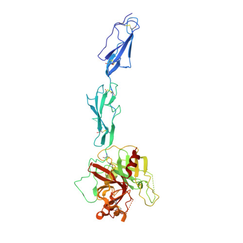
    1GPZ

  • PubMed Abstract: 

    C1r is the modular serine protease (SP) that mediates autolytic activation of C1, the macromolecular complex that triggers the classical pathway of complement. The crystal structure of a mutated, proenzyme form of the catalytic domain of human C1r, comprising the first and second complement control protein modules (CCP1, CCP2) and the SP domain has been solved and refined to 2.9 A resolution. The domain associates as a homodimer with an elongated head-to-tail structure featuring a central opening and involving interactions between the CCP1 module of one monomer and the SP domain of its counterpart. Consequently, the catalytic site of one monomer and the cleavage site of the other are located at opposite ends of the dimer. The structure reveals unusual features in the SP domain and provides strong support for the hypothesis that C1r activation in C1 is triggered by a mechanical stress caused by target recognition that disrupts the CCP1-SP interfaces and allows formation of transient states involving important conformational changes.


  • Organizational Affiliation
    • Laboratoire de Cristallographie et Cristollogénèse des Protéines, Institut de Biologie Structurale Jean-Pierre Ebel, CEA-CNRS-UJF, 41 rue Jules Horowitz, F-38027 Grenoble cedex 1, France.

Macromolecule Content 

  • Total Structure Weight: 92.35 kDa 
  • Atom Count: 5,970 
  • Modeled Residue Count: 737 
  • Deposited Residue Count: 798 
  • Unique protein chains: 1

Macromolecules

Find similar proteins by:|  3D Structure
Entity ID: 1
MoleculeChains  Sequence LengthOrganismDetailsImage
COMPLEMENT C1R COMPONENT
A, B
399Homo sapiensMutation(s): 1 
EC: 3.4.21.41
UniProt & NIH Common Fund Data Resources
Find proteins for P00736 (Homo sapiens)
Explore P00736 
Go to UniProtKB:  P00736
PHAROS:  P00736
Entity Groups
Sequence Clusters30% Identity50% Identity70% Identity90% Identity95% Identity100% Identity
UniProt GroupP00736
Glycosylation
Glycosylation Sites: 2Go to GlyGen: P00736-1
Sequence Annotations
Expand
Reference Sequence

Oligosaccharides

Help  
Entity ID: 2
MoleculeChains Length2D Diagram GlycosylationD Interactions
2-acetamido-2-deoxy-beta-D-glucopyranose-(1-4)-[alpha-L-fucopyranose-(1-6)]2-acetamido-2-deoxy-beta-D-glucopyranose
C
3N-Glycosylation
Glycosylation Resources
GlyTouCan: G21290RB
GlyCosmos: G21290RB
GlyGen: G21290RB
Entity ID: 3
MoleculeChains Length2D Diagram GlycosylationD Interactions
alpha-D-mannopyranose-(1-4)-2-acetamido-2-deoxy-beta-D-glucopyranose-(1-4)-[alpha-L-fucopyranose-(1-6)]2-acetamido-2-deoxy-beta-D-glucopyranose
D
4N-Glycosylation
Glycosylation Resources
GlyTouCan: G47477HI
GlyCosmos: G47477HI
GlyGen: G47477HI

Small Molecules

Ligands 1 Unique
IDChains Name / Formula / InChI Key2D Diagram3D Interactions
NAG

Query on NAG


Download:Ideal Coordinates CCD File
E [auth B]2-acetamido-2-deoxy-beta-D-glucopyranose
C8 H15 N O6
OVRNDRQMDRJTHS-FMDGEEDCSA-N

Experimental Data & Validation

Experimental Data

  • Method: X-RAY DIFFRACTION
  • Resolution: 2.90 Å
  • R-Value Free:  0.290 (Depositor), 0.269 (DCC) 
  • R-Value Work:  0.242 (Depositor), 0.223 (DCC) 
  • R-Value Observed: 0.242 (Depositor) 
Space Group: P 21 21 21
Unit Cell:
Length ( Å )Angle ( ˚ )
a = 99.3α = 90
b = 101.8β = 90
c = 122.4γ = 90
Software Package:
Software NamePurpose
CNSrefinement
MOSFLMdata reduction
CCP4data scaling
AMoREphasing

Structure Validation

View Full Validation Report


Ligand Structure Quality Assessment 


Entry History 

Deposition Data

Revision History  (Full details and data files)

  • Version 1.0: 2002-07-31
    Type: Initial release
  • Version 1.1: 2011-09-28
    Changes: Advisory, Atomic model, Derived calculations, Non-polymer description, Other, Refinement description, Structure summary, Version format compliance
  • Version 1.2: 2017-07-12
    Changes: Advisory
  • Version 1.3: 2019-04-03
    Changes: Data collection, Source and taxonomy
  • Version 1.4: 2019-05-08
    Changes: Data collection, Experimental preparation
  • Version 2.0: 2020-07-29
    Type: Remediation
    Reason: Carbohydrate remediation
    Changes: Advisory, Atomic model, Data collection, Derived calculations, Other, Structure summary
  • Version 2.1: 2023-12-13
    Changes: Advisory, Data collection, Database references, Derived calculations, Refinement description, Structure summary
  • Version 2.2: 2024-11-06
    Changes: Structure summary