1GPQ

Structure of ivy complexed with its target, HEWL


Experimental Data Snapshot

  • Method: X-RAY DIFFRACTION
  • Resolution: 1.60 Å
  • R-Value Free: 0.212 
  • R-Value Work: 0.181 
  • R-Value Observed: 0.181 

wwPDB Validation   3D Report Full Report


This is version 1.3 of the entry. See complete history


Literature

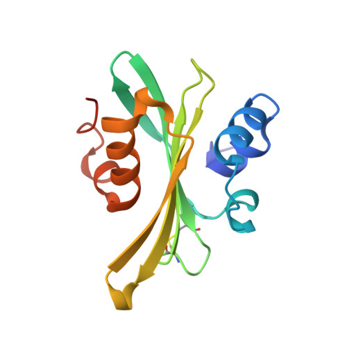
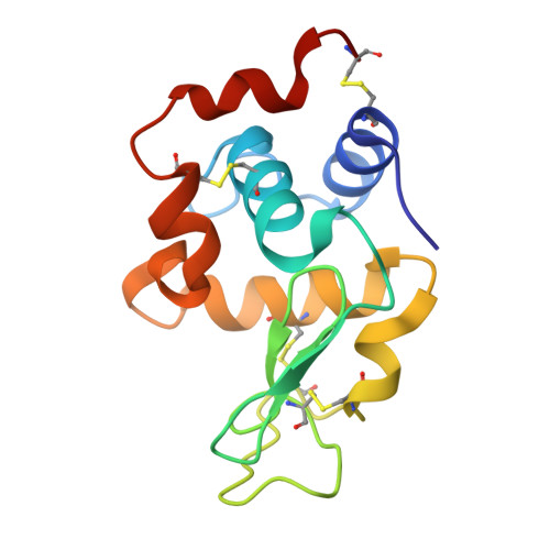
Structure and Evolution of the Ivy Protein Family, Unexpected Lysozyme Inhibitors in Gram-Negative Bacteria.

Abergel, C.Monchois, V.Byrne, D.Chenivesse, S.Lembo, F.Lazzaroni, J.Claverie, J.

(2007) Proc Natl Acad Sci U S A 104: 6394

  • DOI: https://doi.org/10.1073/pnas.0611019104
  • Primary Citation of Related Structures:  
    1GPQ, 1UUZ, 1XS0

  • PubMed Abstract: 

    Part of an ancestral bactericidal system, vertebrate C-type lysozyme targets the peptidoglycan moiety of bacterial cell walls. We report the crystal structure of a protein inhibitor of C-type lysozyme, the Escherichia coli Ivy protein, alone and in complex with hen egg white lysozyme. Ivy exhibits a novel fold in which a protruding five-residue loop appears essential to its inhibitory effect. This feature guided the identification of Ivy orthologues in other Gram-negative bacteria. The structure of the evolutionary distant Pseudomonas aeruginosa Ivy orthologue was also determined in complex with hen egg white lysozyme, and its antilysozyme activity was confirmed. Ivy expression protects porous cell-wall E. coli mutants from the lytic effect of lysozyme, suggesting that it is a response against the permeabilizing effects of the innate vertebrate immune system. As such, Ivy acts as a virulence factor for a number of Gram-negative bacteria-infecting vertebrates.


  • Organizational Affiliation

    Structural and Genomic Information Laboratory, Centre National de la Recherche Scientifique, Unité Propre de Recherche 2589, 13288 Marseille, Cedex 9, France. chantal.abergel@igs.cnrs-mrs.fr


Macromolecules
Find similar proteins by:  (by identity cutoff)  |  3D Structure
Entity ID: 1
MoleculeChains Sequence LengthOrganismDetailsImage
INHIBITOR OF VERTEBRATE LYSOZYME
A, B
135Escherichia coli K-12Mutation(s): 0 
UniProt
Find proteins for P0AD59 (Escherichia coli (strain K12))
Explore P0AD59 
Go to UniProtKB:  P0AD59
Entity Groups  
Sequence Clusters30% Identity50% Identity70% Identity90% Identity95% Identity100% Identity
UniProt GroupP0AD59
Sequence Annotations
Expand
  • Reference Sequence
Find similar proteins by:  (by identity cutoff)  |  3D Structure
Entity ID: 2
MoleculeChains Sequence LengthOrganismDetailsImage
LYSOZYME C
C, D
129Gallus gallusMutation(s): 0 
EC: 3.2.1.17
UniProt
Find proteins for P00698 (Gallus gallus)
Explore P00698 
Go to UniProtKB:  P00698
Entity Groups  
Sequence Clusters30% Identity50% Identity70% Identity90% Identity95% Identity100% Identity
UniProt GroupP00698
Sequence Annotations
Expand
  • Reference Sequence
Experimental Data & Validation

Experimental Data

  • Method: X-RAY DIFFRACTION
  • Resolution: 1.60 Å
  • R-Value Free: 0.212 
  • R-Value Work: 0.181 
  • R-Value Observed: 0.181 
  • Space Group: P 1 21 1
Unit Cell:
Length ( Å )Angle ( ˚ )
a = 55.49α = 90
b = 59.56β = 95.39
c = 69.2γ = 90
Software Package:
Software NamePurpose
DENZOdata reduction
SCALAdata scaling
SOLVEphasing
AMoREphasing
CNSrefinement

Structure Validation

View Full Validation Report


Entry History 

Deposition Data

Revision History  (Full details and data files)

  • Version 1.0: 2003-03-11
    Type: Initial release
  • Version 1.1: 2011-06-23
    Changes: Version format compliance
  • Version 1.2: 2011-07-13
    Changes: Version format compliance
  • Version 1.3: 2023-12-13
    Changes: Data collection, Database references, Other, Refinement description